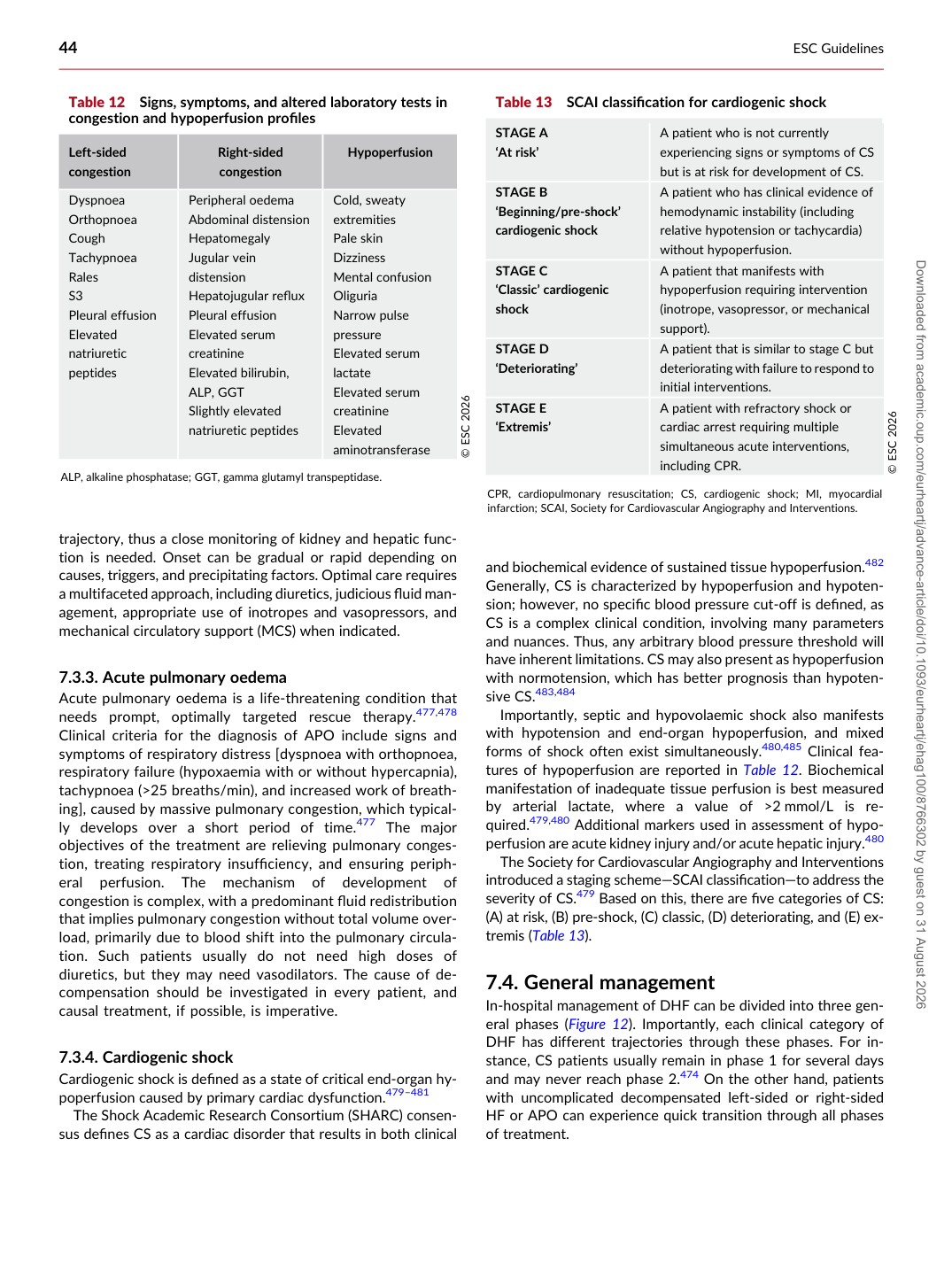
p.44 抽出テキスト
trajectory, thus a close monitoring of kidney and hepatic function is needed. Onset can be gradual or rapid depending on causes, triggers, and precipitating factors. Optimal care requires a multifaceted approach, including diuretics, judicious fluid management, appropriate use of inotropes and vasopressors, and mechanical circulatory support (MCS) when indicated.

7.3.3 Acute pulmonary oedema

Acute pulmonary oedema is a life-threatening condition that needs prompt, optimally targeted rescue therapy.477,478 Clinical criteria for the diagnosis of APO include signs and symptoms of respiratory distress [dyspnoea with orthopnoea, respiratory failure (hypoxaemia with or without hypercapnia), tachypnoea (>25 breaths/min), and increased work of breathing], caused by massive pulmonary congestion, which typically develops over a short period of time.477 The major objectives of the treatment are relieving pulmonary congestion, treating respiratory insufficiency, and ensuring peripheral perfusion. The mechanism of development of congestion is complex, with a predominant fluid redistribution that implies pulmonary congestion without total volume overload, primarily due to blood shift into the pulmonary circulation. Such patients usually do not need high doses of diuretics, but they may need vasodilators. The cause of decompensation should be investigated in every patient, and causal treatment, if possible, is imperative.

7.3.4 Cardiogenic shock

Cardiogenic shock is defined as a state of critical end-organ hypoperfusion caused by primary cardiac dysfunction.479–481 The Shock Academic Research Consortium (SHARC) consensus defines CS as a cardiac disorder that results in both clinical and biochemical evidence of sustained tissue hypoperfusion.482

Generally, CS is characterized by hypoperfusion and hypotension; however, no specific blood pressure cut-off is defined, as CS is a complex clinical condition, involving many parameters and nuances. Thus, any arbitrary blood pressure threshold will have inherent limitations. CS may also present as hypoperfusion with normotension, which has better prognosis than hypotensive CS.483,484 Importantly, septic and hypovolaemic shock also manifests with hypotension and end-organ hypoperfusion, and mixed forms of shock often exist simultaneously.480,485 Clinical features of hypoperfusion are reported in Table 12. Biochemical manifestation of inadequate tissue perfusion is best measured by arterial lactate, where a value of >2 mmol/L is required.479,480 Additional markers used in assessment of hypoperfusion are acute kidney injury and/or acute hepatic injury.480 The Society for Cardiovascular Angiography and Interventions introduced a staging scheme—SCAI classification—to address the severity of CS.479 Based on this, there are five categories of CS: (A) at risk, (B) pre-shock, (C) classic, (D) deteriorating, and (E) extremis (Table 13).

7.4 General management

In-hospital management of DHF can be divided into three general phases (Figure 12). Importantly, each clinical category of DHF has different trajectories through these phases. For instance, CS patients usually remain in phase 1 for several days and may never reach phase 2.474 On the other hand, patients with uncomplicated decompensated left-sided or right-sided HF or APO can experience quick transition through all phases of treatment.

Table 12 Signs, symptoms, and altered laboratory tests in congestion and hypoperfusion profiles

Left-sided congestion Right-sided

congestion Hypoperfusion

Dyspnoea Orthopnoea Cough Tachypnoea Rales S3 Pleural effusion Elevated natriuretic peptides Peripheral oedema Abdominal distension

Hepatomegaly Jugular vein distension Hepatojugular reflux Pleural effusion Elevated serum creatinine Elevated bilirubin, ALP, GGT Slightly elevated natriuretic peptides Cold, sweaty extremities

Pale skin Dizziness Mental confusion Oliguria Narrow pulse pressure Elevated serum lactate Elevated serum creatinine Elevated aminotransferase © ESC 2026

ALP, alkaline phosphatase; GGT, gamma glutamyl transpeptidase. Table 13 SCAI classification for cardiogenic shock

STAGE A ‘At risk’ A patient who is not currently

experiencing signs or symptoms of CS but is at risk for development of CS.

STAGE B ‘Beginning/pre-shock’ cardiogenic shock A patient who has clinical evidence of hemodynamic instability (including

relative hypotension or tachycardia) without hypoperfusion.

STAGE C ‘Classic’ cardiogenic shock A patient that manifests with

hypoperfusion requiring intervention (inotrope, vasopressor, or mechanical support).

STAGE D ‘Deteriorating’ A patient that is similar to stage C but

deteriorating with failure to respond to initial interventions.

STAGE E ‘Extremis’ A patient with refractory shock or cardiac arrest requiring multiple

simultaneous acute interventions, including CPR. © ESC 2026

CPR, cardiopulmonary resuscitation; CS, cardiogenic shock; MI, myocardial infarction; SCAI, Society for Cardiovascular Angiography and Interventions. Downloaded from academic.oup.com/eurheartj/advance-article/doi/10.1093/eurheartj/ehag100/8766302 by guest on 31 August 2026