p.30 抽出テキスト
of arrhythmia-associated HF is important in patients with HFrEF, in whom a rhythm strategy for AF may potentially be beneficial. The diagnostic work-up may include an ECG and ambulatory ECG to characterize the electrical subtype, and CMR imaging to characterize the morphological/functional subtype. Arrhythmias may be the sole cause of HFrEF or merely a bystander in the context of other causes of HF. Heart failure associated with abnormal cardiac conduction is defined as LV dysfunction due to abnormal electrical conduction and should be suspected in patients with LV dysfunction and a QRS duration ≥130 ms, without evidence of other causes for HF.199 It should also be considered in patients with implanted pacemakers or implantable cardioverter-defibrillators (ICDs) delivering RV pacing (see Section 6.2.2.2).200–203 The diagnosis is a diagnosis of elimination, and it may only be reached retrospectively, if LV function improves following cardiac resynchronization therapy (CRT) implantation.204

In terms of likelihood of response, there is a spectrum. At one end, abnormal cardiac conduction may be the sole culprit, whereas at the other, it may be a bystander in the context of HF due to other causes (Figure 9). CMR may be useful in identifying myocardial scar and morphological abnormalities pointing to other aetiologies of HF. Abnormal cardiac conduction, as the sole cause of HFrEF, is most likely in patients with HFrEF, LBBB, wide QRS duration, with no myocardial scarring on CMR and in patients with RV pacing-induced HF.199

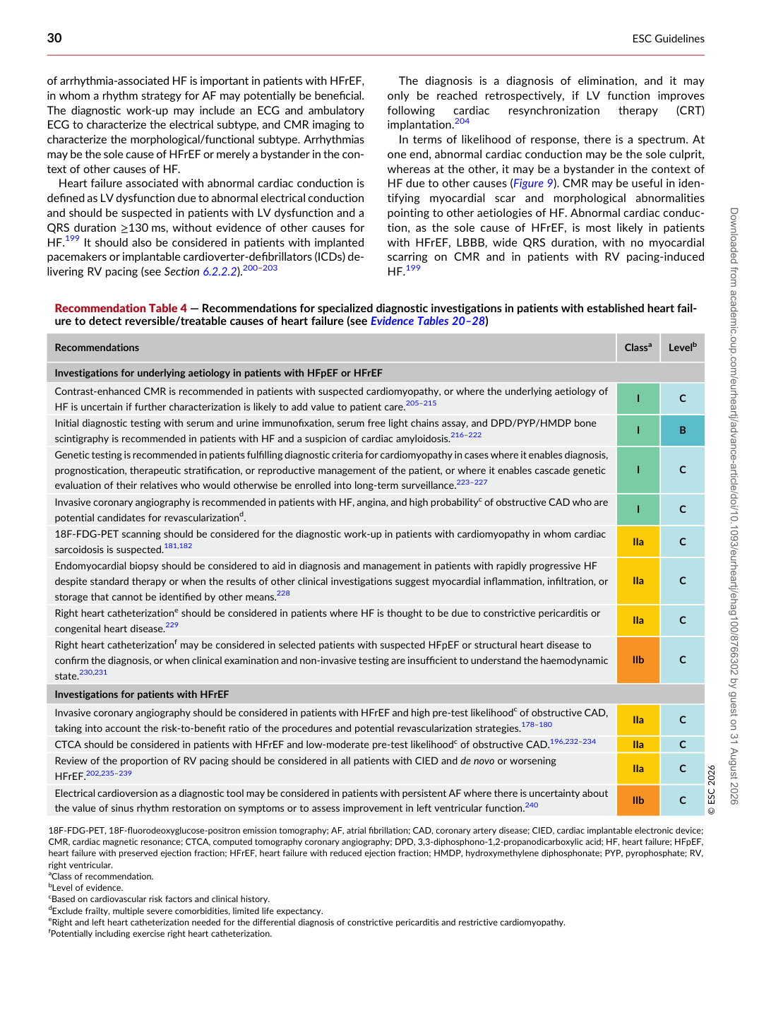
Recommendation Table 4 — Recommendations for specialized diagnostic investigations in patients with established heart failure to detect reversible/treatable causes of heart failure (see Evidence Tables 20–28)

Recommendations Classa Levelb

Investigations for underlying aetiology in patients with HFpEF or HFrEF Contrast-enhanced CMR is recommended in patients with suspected cardiomyopathy, or where the underlying aetiology of HF is uncertain if further characterization is likely to add value to patient care.205–215 I C Initial diagnostic testing with serum and urine immunofixation, serum free light chains assay, and DPD/PYP/HMDP bone scintigraphy is recommended in patients with HF and a suspicion of cardiac amyloidosis.216–222 I B Genetic testing is recommended in patients fulfilling diagnostic criteria for cardiomyopathy in cases where it enables diagnosis, prognostication, therapeutic stratification, or reproductive management of the patient, or where it enables cascade genetic evaluation of their relatives who would otherwise be enrolled into long-term surveillance.223–227 I C Invasive coronary angiography is recommended in patients with HF, angina, and high probabilityc of obstructive CAD who are potential candidates for revascularizationd. I C 18F-FDG-PET scanning should be considered for the diagnostic work-up in patients with cardiomyopathy in whom cardiac sarcoidosis is suspected.181,182 IIa C Endomyocardial biopsy should be considered to aid in diagnosis and management in patients with rapidly progressive HF despite standard therapy or when the results of other clinical investigations suggest myocardial inflammation, infiltration, or storage that cannot be identified by other means.228 IIa C Right heart catheterizatione should be considered in patients where HF is thought to be due to constrictive pericarditis or congenital heart disease.229 IIa C Right heart catheterizationf may be considered in selected patients with suspected HFpEF or structural heart disease to confirm the diagnosis, or when clinical examination and non-invasive testing are insufficient to understand the haemodynamic state.230,231 IIb C Investigations for patients with HFrEF Invasive coronary angiography should be considered in patients with HFrEF and high pre-test likelihoodc of obstructive CAD, taking into account the risk-to-benefit ratio of the procedures and potential revascularization strategies.178–180 IIa C CTCA should be considered in patients with HFrEF and low-moderate pre-test likelihoodc of obstructive CAD.196,232–234 IIa C Review of the proportion of RV pacing should be considered in all patients with CIED and de novo or worsening HFrEF.202,235–239 IIa C Electrical cardioversion as a diagnostic tool may be considered in patients with persistent AF where there is uncertainty about the value of sinus rhythm restoration on symptoms or to assess improvement in left ventricular function.240 IIb C © ESC 2026 18F-FDG-PET, 18F-fluorodeoxyglucose-positron emission tomography; AF, atrial fibrillation; CAD, coronary artery disease; CIED, cardiac implantable electronic device; CMR, cardiac magnetic resonance; CTCA, computed tomography coronary angiography; DPD, 3,3-diphosphono-1,2-propanodicarboxylic acid; HF, heart failure; HFpEF, heart failure with preserved ejection fraction; HFrEF, heart failure with reduced ejection fraction; HMDP, hydroxymethylene diphosphonate; PYP, pyrophosphate; RV, right ventricular. aClass of recommendation. bLevel of evidence. cBased on cardiovascular risk factors and clinical history. dExclude frailty, multiple severe comorbidities, limited life expectancy. eRight and left heart catheterization needed for the differential diagnosis of constrictive pericarditis and restrictive cardiomyopathy. fPotentially including exercise right heart catheterization. Downloaded from academic.oup.com/eurheartj/advance-article/doi/10.1093/eurheartj/ehag100/8766302 by guest on 31 August 2026