p.29 抽出テキスト
testing may be needed (see Supplementary data online, Table S2). Advances in multimodal imaging, including CMR, stress testing, and computed tomography (CT), have significantly enhanced our capacity to comprehensively assess HF, offering complementary information that is crucial in complex cases.183–185 CMR is considered the gold standard for assessing LV volumes and mass, owing to its high spatial resolution and reproducibility. Late gadolinium enhancement (LGE) provides detailed insights into fibrosis and scarring and is useful in distinguishing ischaemic from non-ischaemic aetiology as well as certain cardiomyopathies.124 CMR myocardial tissue characterization with parametric mapping [T1, T2, T2* (longitudinal, transverse, and observed transverse relaxation times), LGE and extracellular volume (ECV) expansion] enables classification of cardiomyopathies124,186–193 and of cardiac amyloid.194 An overview of typical CMR findings in cardiomyopathies can be found in the 2023 ESC Guidelines for the management of cardiomyopathies.124 Description of other specific imaging modalities is provided in the Sections on specific comorbidities.

5.3.2 Investigations for coronary artery disease

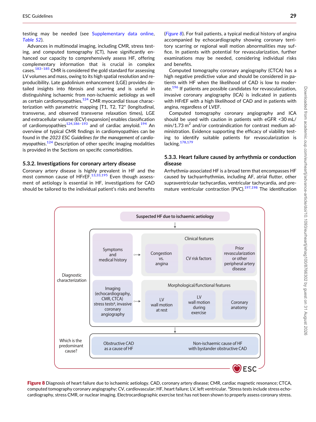
Coronary artery disease is highly prevalent in HF and the most common cause of HFrEF.13,33,195 Even though assessment of aetiology is essential in HF, investigations for CAD should be tailored to the individual patient’s risks and benefits (Figure 8). For frail patients, a typical medical history of angina accompanied by echocardiography showing coronary territory scarring or regional wall motion abnormalities may suffice. In patients with potential for revascularization, further examinations may be needed, considering individual risks and benefits.

Computed tomography coronary angiography (CTCA) has a high negative predictive value and should be considered in patients with HF when the likelihood of CAD is low to moderate.196 If patients are possible candidates for revascularization, invasive coronary angiography (ICA) is indicated in patients with HFrEF with a high likelihood of CAD and in patients with angina, regardless of LVEF. Computed tomography coronary angiography and ICA should be used with caution in patients with eGFR <30 mL/ min/1.73 m2 and/or contraindication for contrast medium administration. Evidence supporting the efficacy of viability testing to identify suitable patients for revascularization is lacking.178,179

5.3.3 Heart failure caused by arrhythmia or conduction

disease Arrhythmia-associated HF is a broad term that encompasses HF caused by tachyarrhythmias, including AF, atrial flutter, other supraventricular tachycardias, ventricular tachycardia, and premature ventricular contraction (PVC).197,198 The identification

Figure 8 Diagnosis of heart failure due to ischaemic aetiology. CAD, coronary artery disease; CMR, cardiac magnetic resonance; CTCA, computed tomography coronary angiography; CV, cardiovascular; HF, heart failure; LV, left ventricular. aStress tests include stress echocardiography, stress CMR, or nuclear imaging. Electrocardiographic exercise test has not been shown to properly assess coronary stress. Downloaded from academic.oup.com/eurheartj/advance-article/doi/10.1093/eurheartj/ehag100/8766302 by guest on 31 August 2026