p.25 抽出テキスト
diagnostic pathway, especially if echocardiography is not readily available. However, many factors may cause false positive or false negative results, which should be considered when evaluating natriuretic peptide (Table 9). LVEF is a continuous measure and may differ between methods used as well as sex. For this reason, a precise value is only given as guidance. Aetiology assessment is important, as it may result in finding potential treatable and/or reversible causes. The major aetiologies of HFrEF are illustrated in Figure 5.

5.2.2 Heart failure with preserved ejection fraction

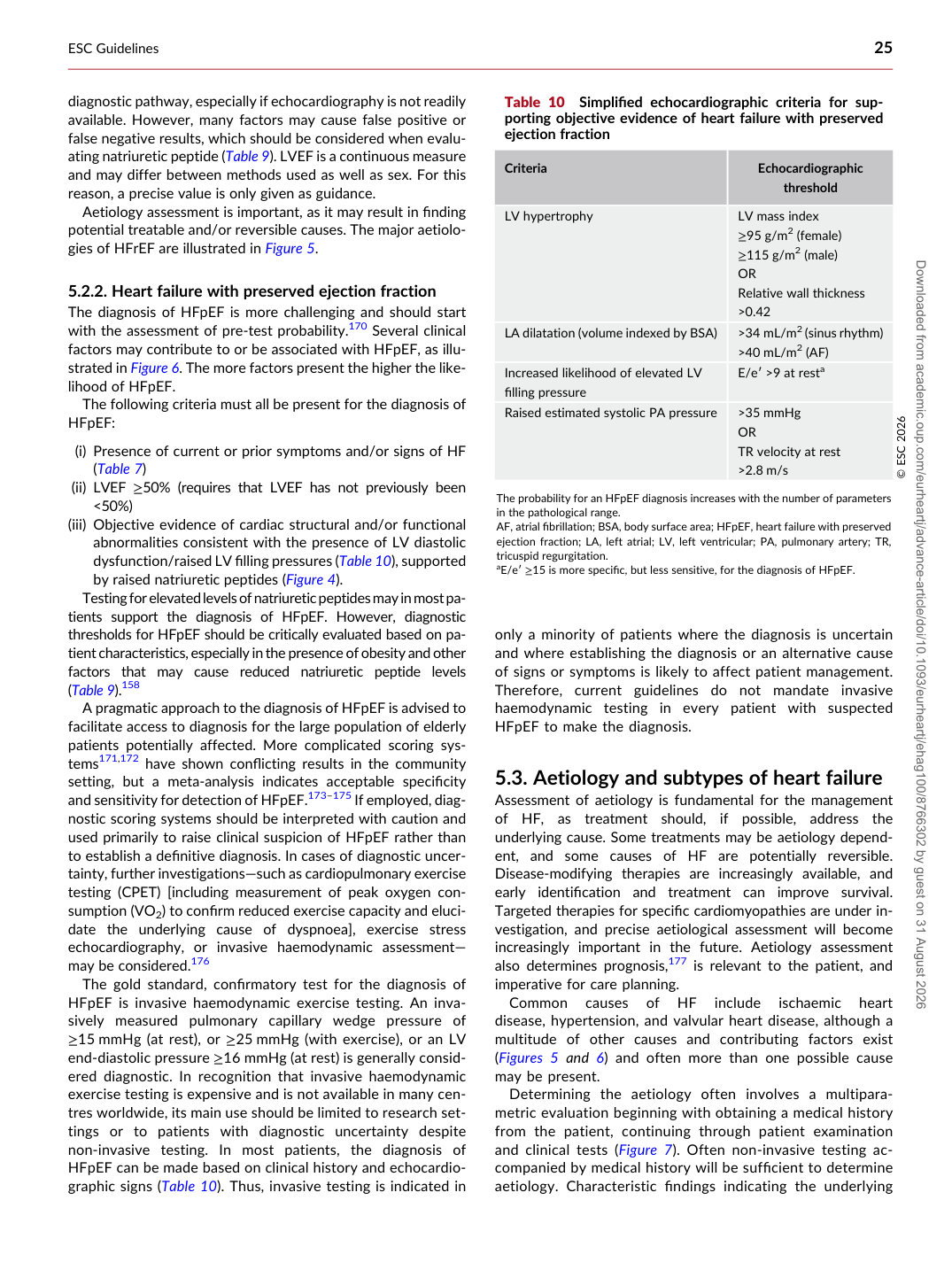
The diagnosis of HFpEF is more challenging and should start with the assessment of pre-test probability.170 Several clinical factors may contribute to or be associated with HFpEF, as illustrated in Figure 6. The more factors present the higher the likelihood of HFpEF. The following criteria must all be present for the diagnosis of HFpEF: (i) Presence of current or prior symptoms and/or signs of HF (Table 7) (ii) LVEF ≥50% (requires that LVEF has not previously been <50%) (iii) Objective evidence of cardiac structural and/or functional abnormalities consistent with the presence of LV diastolic dysfunction/raised LV filling pressures (Table 10), supported by raised natriuretic peptides (Figure 4). Testing for elevated levels of natriuretic peptides may in most patients support the diagnosis of HFpEF. However, diagnostic thresholds for HFpEF should be critically evaluated based on patient characteristics, especially in the presence of obesity and other factors that may cause reduced natriuretic peptide levels (Table 9).158 A pragmatic approach to the diagnosis of HFpEF is advised to facilitate access to diagnosis for the large population of elderly patients potentially affected. More complicated scoring systems171,172 have shown conflicting results in the community setting, but a meta-analysis indicates acceptable specificity and sensitivity for detection of HFpEF.173–175 If employed, diagnostic scoring systems should be interpreted with caution and used primarily to raise clinical suspicion of HFpEF rather than to establish a definitive diagnosis. In cases of diagnostic uncertainty, further investigations—such as cardiopulmonary exercise testing (CPET) [including measurement of peak oxygen consumption (VO2) to confirm reduced exercise capacity and elucidate the underlying cause of dyspnoea], exercise stress echocardiography, or invasive haemodynamic assessment— may be considered.176 The gold standard, confirmatory test for the diagnosis of HFpEF is invasive haemodynamic exercise testing. An invasively measured pulmonary capillary wedge pressure of ≥15 mmHg (at rest), or ≥25 mmHg (with exercise), or an LV end-diastolic pressure ≥16 mmHg (at rest) is generally considered diagnostic. In recognition that invasive haemodynamic exercise testing is expensive and is not available in many centres worldwide, its main use should be limited to research settings or to patients with diagnostic uncertainty despite non-invasive testing. In most patients, the diagnosis of HFpEF can be made based on clinical history and echocardiographic signs (Table 10). Thus, invasive testing is indicated in only a minority of patients where the diagnosis is uncertain and where establishing the diagnosis or an alternative cause of signs or symptoms is likely to affect patient management. Therefore, current guidelines do not mandate invasive haemodynamic testing in every patient with suspected HFpEF to make the diagnosis.

5.3 Aetiology and subtypes of heart failure

Assessment of aetiology is fundamental for the management of HF, as treatment should, if possible, address the underlying cause. Some treatments may be aetiology dependent, and some causes of HF are potentially reversible. Disease-modifying therapies are increasingly available, and early identification and treatment can improve survival. Targeted therapies for specific cardiomyopathies are under investigation, and precise aetiological assessment will become increasingly important in the future. Aetiology assessment also determines prognosis,177 is relevant to the patient, and imperative for care planning. Common causes of HF include ischaemic heart disease, hypertension, and valvular heart disease, although a multitude of other causes and contributing factors exist (Figures 5 and 6) and often more than one possible cause may be present. Determining the aetiology often involves a multiparametric evaluation beginning with obtaining a medical history from the patient, continuing through patient examination and clinical tests (Figure 7). Often non-invasive testing accompanied by medical history will be sufficient to determine aetiology. Characteristic findings indicating the underlying

Table 10 Simplified echocardiographic criteria for supporting objective evidence of heart failure with preserved ejection fraction

Criteria Echocardiographic

threshold

LV hypertrophy LV mass index

≥95 g/m2 (female) ≥115 g/m2 (male) OR Relative wall thickness >0.42

LA dilatation (volume indexed by BSA) >34 mL/m2 (sinus rhythm) >40 mL/m2 (AF) Increased likelihood of elevated LV filling pressure E/e′ >9 at resta Raised estimated systolic PA pressure >35 mmHg OR

TR velocity at rest >2.8 m/s © ESC 2026

The probability for an HFpEF diagnosis increases with the number of parameters in the pathological range. AF, atrial fibrillation; BSA, body surface area; HFpEF, heart failure with preserved ejection fraction; LA, left atrial; LV, left ventricular; PA, pulmonary artery; TR, tricuspid regurgitation. aE/e′ ≥15 is more specific, but less sensitive, for the diagnosis of HFpEF. Downloaded from academic.oup.com/eurheartj/advance-article/doi/10.1093/eurheartj/ehag100/8766302 by guest on 31 August 2026