p.63 抽出テキスト
14.4 Management of prosthetic valve

dysfunction and complications Prosthetic valve dysfunction can occur due to intrinsic permanent changes to the prosthetic valve (defined as SVD) or non-structural valve dysfunction resulting from any abnormality not intrinsic to the prosthetic valve itself. Valve thrombosis and endocarditis are considered separate entities due to their specific presentation and management, but may both result in SVD.

14.4.1 Structural valve deterioration

Structural valve deterioration has been defined by several consensus documents812,895,896 and includes wear and tear, leaflet disruption, leaflet fibrosis or calcification, and stent or strut fracture or deformation. BHV (transcatheter or surgical) SVD is more frequent than MHV SVD. The incidence of SVD may be underestimated by the simple analysis of patients with valve-related deaths or those undergoing reintervention. To ensure timely diagnosis, serial measurement should be performed and compared with the TTE performed at discharge, or within 1–3 months after valve implantation.

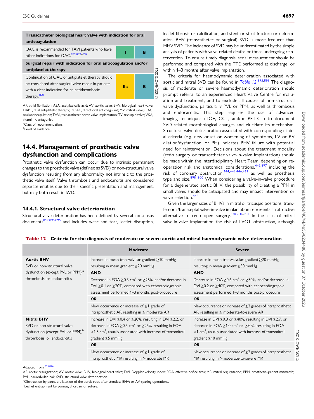
The criteria for haemodynamic deterioration associated with aortic and mitral SVD can be found in Table 12.895,896 The diagnosis of moderate or severe haemodynamic deterioration should prompt referral to an experienced Heart Valve Centre for evaluation and treatment, and to exclude all causes of non-structural valve dysfunction, particularly PVL or PPM, as well as thrombosis and endocarditis. This step requires the use of advanced imaging techniques (TOE, CCT, and/or PET-CT) to document SVD-related morphological changes and elucidate its mechanism. Structural valve deterioration associated with corresponding clinical criteria (e.g. new onset or worsening of symptoms, LV or RV dilation/dysfunction, or PH) indicates BHV failure with potential need for reintervention. Decisions about the treatment modality (redo surgery or transcatheter valve-in-valve implantation) should be made within the interdisciplinary Heart Team, depending on reoperation risk and anatomical considerations,445,897 including the risk of coronary obstruction,144,442,446,461 as well as prosthesis type and size.898–900 When considering a valve-in-valve procedure for a degenerated aortic BHV, the possibility of creating a PPM in small valves should be anticipated and may impact intervention or valve selection.448 Given the larger sizes of BHVs in mitral or tricuspid positions, transfemoral/transseptal valve-in-valve implantation represents an attractive alternative to redo open surgery.570,900–903 In the case of mitral valve-in-valve implantation the risk of LVOT obstruction, although

Transcatheter biological heart valve with indication for oral anticoagulation OAC is recommended for TAVI patients who have other indications for OAC.879,892–894 I B Surgical repair with indication for oral anticoagulation and/or antiplatelet therapy Continuation of OAC or antiplatelet therapy should be considered after surgical valve repair in patients with a clear indication for an antithrombotic therapy.890 IIa B © ESC/EACTS 2025

AF, atrial fibrillation; ASA, acetylsalicylic acid; AV, aortic valve; BHV, biological heart valve; DAPT, dual antiplatelet therapy; DOAC, direct oral anticoagulant; MV, mitral valve; OAC, oral anticoagulation; TAVI, transcatheter aortic valve implantation; TV, tricuspid valve; VKA, vitamin K antagonist. aClass of recommendation. bLevel of evidence.

Table 12 Criteria for the diagnosis of moderate or severe aortic and mitral haemodynamic valve deterioration

Moderate Severe

Aortic BHV SVD or non-structural valve dysfunction (except PVL or PPM),a thrombosis, or endocarditis Increase in mean transvalvular gradient ≥10 mmHg

resulting in mean gradient ≥20 mmHg Increase in mean transvalvular gradient ≥20 mmHg

resulting in mean gradient ≥30 mmHg

AND AND Decrease in EOA ≥0.3 cm2 or ≥25%, and/or decrease in DVI ≥0.1 or ≥20%, compared with echocardiographic assessment performed 1–3 months post-procedure Decrease in EOA ≥0.6 cm2 or ≥50%, and/or decrease in DVI ≥0.2 or ≥40%, compared with echocardiographic

assessment performed 1–3 months post-procedure

OR OR New occurrence or increase of ≥1 grade of intraprosthetic AR resulting in ≥ moderate AR New occurrence or increase of ≥2 grades of intraprosthetic AR resulting in ≥ moderate-to-severe AR

Mitral BHV SVD or non-structural valve dysfunction (except PVL or PPM),b thrombosis, or endocarditis Increase in DVI ≥0.4 or ≥20%, resulting in DVI ≥2.2, or decrease in EOA ≥0.5 cm2 or ≥25%, resulting in EOA

<1.5 cm2, usually associated with increase of transmitral gradient ≥5 mmHg Increase in DVI ≥0.8 or ≥40%, resulting in DVI ≥2.7, or decrease in EOA ≥1.0 cm2 or ≥50%, resulting in EOA

<1 cm2, usually associated with increase of transmitral gradient ≥10 mmHg

OR OR New occurrence or increase of ≥1 grade of intraprosthetic MR resulting in ≥moderate MR New occurrence or increase of ≥2 grades of intraprosthetic

MR resulting in ≥moderate-to-severe MR © ESC/EACTS 2025

Adapted from 895,896. AR, aortic regurgitation; AV, aortic valve; BHV, biological heart valve; DVI, Doppler velocity index; EOA, effective orifice area; MR, mitral regurgitation; PPM, prosthesis–patient mismatch; PVL, paravalvular leak; SVD, structural valve deterioration. aObstruction by pannus; dilatation of the aortic root after stentless BHV; or AV-sparing operations. bLeaflet entrapment by pannus, chordae, or suture.

ESC Guidelines                                                                                                                                                                                          4697

Downloaded from academic.oup.com/eurheartj/article/46/44/4635/8234488 by guest on 07 October 2026