p.48 抽出テキスト
surgical MV repair or replacement with extensive decalcification and patch reconstruction of the annulus can be performed in selected patients at experienced centres (e.g. young patients post-chest radiation), where mortality rates of <5% have been reported.677–679 Degenerative MS is not amenable to PMC because commissural fusion is absent. In symptomatic high-risk patients with suitable anatomy, transcatheter implantation of a TAVI prosthesis in the mitral position is feasible but associated with frequent complications, including LVOT obstruction, valve embolization, stroke, and haemolysis due to PVL. Open surgical valve replacement via the LA with a TAVI device is an alternative that allows complete anterior leaflet removal;680 however, mortality remains high. The use of dedicated TMVI devices is therefore encouraged because it appears to be safer.542,681 Heart Team evaluation should guide the choice of treatment avoiding futility, because mortality remains high, even after successful treatment (10%–30% within 1 year).

11 Tricuspid regurgitation

11.1 Prevalence and aetiology

TR is a common echocardiographic finding in the general population, with higher prevalence in women and older patients. Trivial or mild TR is mostly a benign condition. Significant TR (≥ moderate) has a reported age- and sex-adjusted prevalence of 0.55% (4% in people aged ≥75 years).682 Severe TR is associated with increased risk of death and HF, independent of comorbidities, ventricular function, and pulmonary pressures.683–686 Only 8%–10% of patients with TR present with clear anatomical abnormalities of the TV apparatus (primary TR), which can be due to infective endocarditis, RHD, carcinoid syndrome, congenital abnormalities (e.g. Ebstein’s anomaly), chest radiation, or myxomatous disease, as well as trauma or iatrogenic valve damage (e.g. after endomyocardial biopsy).682 Cardiac implantable electronic device (CIED)-related TR represents a separate entity requiring a specific diagnostic approach and management.687 In patients with a CIED, diagnostic efforts should be made to clarify if the lead is the cause of TR (CIED-related TR) or incidental (CIED-associated TR).688 In patients with secondary TR, TV leaflets are structurally normal and regurgitation is caused by annular dilatation and/or leaflet tethering due to RA dilatation, and/or RV dilation and dysfunction. Based on the main morphological and haemodynamic characteristics, two phenotypes of secondary TR have been proposed:689 (i) atrial secondary TR, mainly due to AF and characterized by the absence of significant leaflet tethering, but with marked RA and annular dilatation along with preserved RV size/function, pulmonary pressure, and LV function; and (ii) ventricular secondary TR, due to annular dilatation and leaflet tethering as a consequence of left-sided ventricular or valvular disease (post-capillary PH), pre-capillary PH, or primary RV cardiomyopathy/ischaemia (also after left-sided valve surgery).690 At an advanced disease stage, these two phenotypes may no longer be distinguishable, and therefore early characterization is key to determine outcome.691,692 Evidence of an impact on patient management is currently lacking; therefore, current recommendations for intervention consider mainly primary vs secondary TR.

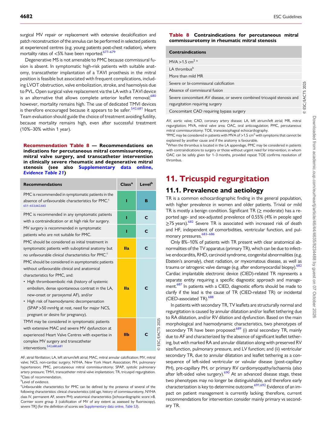
Recommendation Table 8 — Recommendations on indications for percutaneous mitral commissurotomy, mitral valve surgery, and transcatheter intervention in clinically severe rheumatic and degenerative mitral stenosis (see also Supplementary data online, Evidence Table 21)

Recommendations Classa Levelb

PMC is recommended in symptomatic patients in the absence of unfavourable characteristics for PMC.c

651–653,662,665 I B

PMC is recommended in any symptomatic patients with a contraindication or at high risk for surgery. I C MV surgery is recommended in symptomatic patients who are not suitable for PMC. I C PMC should be considered as initial treatment in symptomatic patients with suboptimal anatomy but no unfavourable clinical characteristics for PMC.c IIa C PMC should be considered in asymptomatic patients without unfavourable clinical and anatomical characteristics for PMC, and: • High thromboembolic risk (history of systemic

embolism, dense spontaneous contrast in the LA, new-onset or paroxysmal AF), and/or • High risk of haemodynamic decompensation

(SPAP >50 mmHg at rest, need for major NCS, pregnant or desire for pregnancy). IIa C TMVI may be considered in symptomatic patients with extensive MAC and severe MV dysfunction at experienced Heart Valve Centres with expertise in complex MV surgery and transcatheter interventions.542,680,681 IIb C © ESC/EACTS 2025

AF, atrial fibrillation; LA, left atrium/left atrial; MAC, mitral annular calcification; MV, mitral valve; NCS, non-cardiac surgery; NYHA, New York Heart Association; PH, pulmonary hypertension; PMC, percutaneous mitral commissurotomy; SPAP, systolic pulmonary artery pressure; TMVI, transcatheter mitral valve implantation; TR, tricuspid regurgitation. aClass of recommendation. bLevel of evidence. cUnfavourable characteristics for PMC can be defined by the presence of several of the following characteristics: clinical characteristics (old age, history of commissurotomy, NYHA class IV, permanent AF, severe PH); anatomical characteristics [echocardiographic score >8, Cormier score group 3 (calcification of MV of any extent as assessed by fluoroscopy), severe TR] (for the definition of scores see Supplementary data online, Table S3). Table 8 Contraindications for percutaneous mitral commissurotomy in rheumatic mitral stenosis

Contraindications MVA >1.5 cm2 a LA thrombusb More than mild MR Severe or bi-commissural calcification Absence of commissural fusion Severe concomitant AV disease, or severe combined tricuspid stenosis and regurgitation requiring surgery Concomitant CAD requiring bypass surgery © ESC/EACTS 2025

AV, aortic valve; CAD, coronary artery disease; LA, left atrium/left atrial; MR, mitral regurgitation; MVA, mitral valve area; OAC, oral anticoagulation; PMC, percutaneous mitral commissurotomy; TOE, transoesophageal echocardiography. aPMC may be considered in patients with MVA of >1.5 cm2 with symptoms that cannot be explained by another cause and if the anatomy is favourable. bWhen the thrombus is located in the LA appendage, PMC may be considered in patients with contraindications to surgery or those without urgent need for intervention, in whom OAC can be safely given for 1–3 months, provided repeat TOE confirms resolution of thrombus. Downloaded from academic.oup.com/eurheartj/article/46/44/4635/8234488 by guest on 07 October 2026