p.41 抽出テキスト
9.1.5 Follow-up

Asymptomatic patients with severe MR not fulfilling the criteria for an intervention, and with documented preserved exercise capacity, should undergo clinical and echocardiographic follow-up twice per year (watchful waiting strategy), ideally in the setting of a Heart Valve Clinic.568 Follow-up may also include serial measurements of BNP levels, electrocardiogram (ECG) and/or Holter monitoring, and (in selected cases) exercise echocardiography and CMR to confirm MR severity, and cardiac chamber function and dimensions.46 Asymptomatic patients with moderate MR and preserved LV function can be followed on a yearly basis with echocardiographic assessment every 1 or 2 years. The frequency of follow-up after an intervention depends on the type of procedure performed. Very good long-term durability of MV repair in PMR due to valve prolapse with a low recurrence rate has been reported at experienced centres with freedom from moderate or severe MR of 87.5% at 20 years.533,559 Serial clinical and echocardiographic follow-up in patients without pre-operative LV dysfunction or rhythm abnormalities every 2–3 years thereafter is sufficient. Patients with atrial or ventricular arrhythmias possibly related to MV disease should be further evaluated using continuous ECG monitoring. Patients with recurrent MR after surgical ring annuloplasty frequently undergo repeat surgery (usually MV replacement), while transcatheter alternatives are reserved for selected high-risk patients because of the risk of increased gradients (M-TEER), PVL, and LVOT obstruction (mitral valve-in-ring procedures).569,570 In patients undergoing MV replacement, closer follow-up on a yearly basis is required due to the risk of prosthetic valve dysfunction or SVD (see Section 14.4). Following TEER, reported rates of residual MR and increased transmitral gradients are higher than after surgical repair, suggesting that yearly follow-up is appropriate.571 Although rare, the need for surgical treatment after failed TEER is associated with increased peri-operative mortality and low valve repair rates,541,572 while transcatheter solutions to detach TEER implants and replace the valve have been described in few cases.573,574

9.2 Secondary mitral regurgitation

9.2.1. Prevalence and aetiology Secondary mitral regurgitation is present when the MV structure appears grossly normal but the MV is nonetheless incompetent, due to alterations in LV and LA geometry, dyssynchrony, and imbalances between MV closing and tethering forces.575 The prevalence of severe SMR in patients with chronic HF is ∼10% and higher in patients with reduced vs preserved LVEF (25% vs 4%).576 Secondary mitral regurgitation can be classified as atrial or ventricular with different pathophysiological and morphological characteristics, as well as contrasting prognostic and therapeutic implications.577 Ventricular SMR is more common and associated with worse long-term prognosis.513,578–580 Dilated or ischaemic cardiomyopathy are the most frequent causes of severe ventricular SMR. Acute HF exacerbation may occur in patients with chronic HF due to a renewed ischaemic event, arrhythmia, infection, or volume overload. Atrial SMR is due to pure mitral annular dilation and is observed in patients with long-standing AF and/or HFpEF.581 Factors predisposing to atrial SMR include age ≥65 years, female sex, LA dilatation, and diastolic dysfunction.582 From a morphological point of view, ventricular SMR is characterized by leaflet tethering and restricted motion combined with annular dilation, while annulus enlargement and flattening leading to planar coaptation are predominantly observed in atrial SMR. The prevalence of atrial SMR has been underestimated in the past and was occasionally misclassified as PMR due to pseudo-prolapse with leaflet tethering in advanced stages.

9.2.2 Evaluation

Echocardiographic criteria to define significant SMR according to aetiology are reported in Figure 10. Importantly, SMR assessment should be performed after optimization of medical therapy and in a euvolaemic and normotensive state. When quantifying EROA and RVol in SMR, lower thresholds may apply to define severe regurgitation because of the potential elliptical regurgitant orifice and/or the low-flow state. An EROA of ≥30 mm2 and/or an RVol of ≥45 mL has been identified as having a significant impact on outcomes,45 with prognosis improved after treatment.583,584 Cardiac magnetic resonance is used to confirm SMR severity and assess cardiac chamber function and dimensions. The extent of myocardial fibrosis, as assessed with CMR, has been associated with poor prognosis.585 Owing to the dynamic nature of SMR, exercise echocardiography may help to identify patients with severe SMR when values at rest are inconclusive.45

9.2.3 Definition of atrial secondary mitral

regurgitation Characteristics distinguishing between atrial and ventricular SMR are displayed in Figure 12.

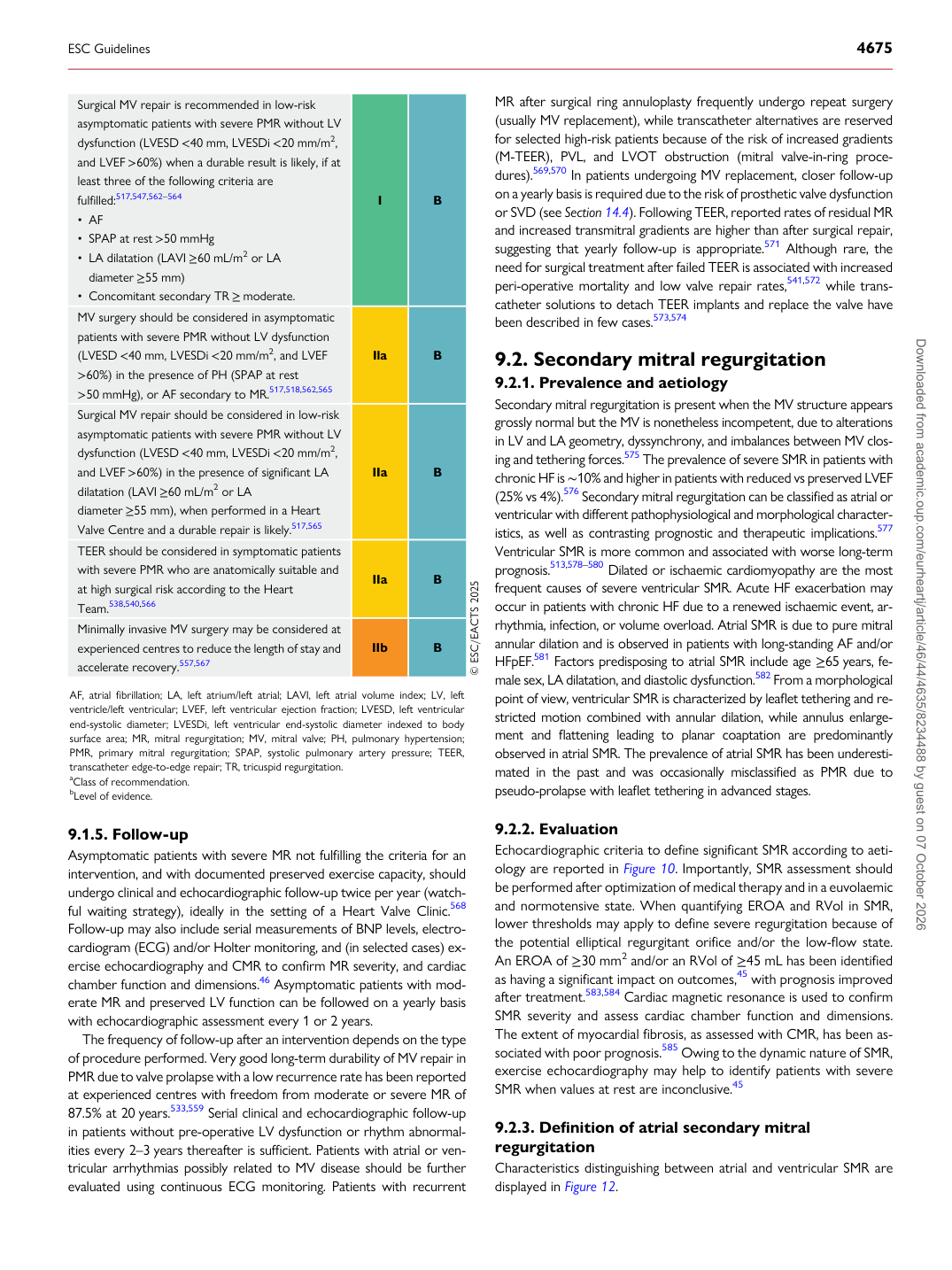
Surgical MV repair is recommended in low-risk asymptomatic patients with severe PMR without LV dysfunction (LVESD <40 mm, LVESDi <20 mm/m2, and LVEF >60%) when a durable result is likely, if at least three of the following criteria are fulfilled:517,547,562–564 • AF • SPAP at rest >50 mmHg • LA dilatation (LAVI ≥60 mL/m2 or LA diameter ≥55 mm) • Concomitant secondary TR ≥ moderate. I B MV surgery should be considered in asymptomatic patients with severe PMR without LV dysfunction (LVESD <40 mm, LVESDi <20 mm/m2, and LVEF >60%) in the presence of PH (SPAP at rest >50 mmHg), or AF secondary to MR.517,518,562,565 Ila B Surgical MV repair should be considered in low-risk asymptomatic patients with severe PMR without LV dysfunction (LVESD <40 mm, LVESDi <20 mm/m2, and LVEF >60%) in the presence of significant LA dilatation (LAVI ≥60 mL/m2 or LA diameter ≥55 mm), when performed in a Heart Valve Centre and a durable repair is likely.517,565 IIa B TEER should be considered in symptomatic patients with severe PMR who are anatomically suitable and at high surgical risk according to the Heart Team.538,540,566 IIa B Minimally invasive MV surgery may be considered at experienced centres to reduce the length of stay and accelerate recovery.557,567 IIb B © ESC/EACTS 2025

AF, atrial fibrillation; LA, left atrium/left atrial; LAVI, left atrial volume index; LV, left ventricle/left ventricular; LVEF, left ventricular ejection fraction; LVESD, left ventricular end-systolic diameter; LVESDi, left ventricular end-systolic diameter indexed to body surface area; MR, mitral regurgitation; MV, mitral valve; PH, pulmonary hypertension; PMR, primary mitral regurgitation; SPAP, systolic pulmonary artery pressure; TEER, transcatheter edge-to-edge repair; TR, tricuspid regurgitation. aClass of recommendation. bLevel of evidence.

ESC Guidelines                                                                                                                                                                                          4675

Downloaded from academic.oup.com/eurheartj/article/46/44/4635/8234488 by guest on 07 October 2026