p.37 抽出テキスト
8.6 Follow-up

The rate of progression of AS varies widely and asymptomatic patients, their family, and medical caregivers need careful education, with particular emphasis on the importance of regular follow-up (ideally at a Heart Valve Centre)14 and prompt reporting of symptoms. Asymptomatic patients with severe AS should be followed up at least every 6 months to allow detection of early symptoms (using exercise testing if complaints are inconclusive) and any change in echocardiographic parameters (particularly LVEF). Serial measurements of natriuretic peptides can provide additional useful information regarding the timing of treatment. Younger patients with mild AS and no significant leaflet calcification may be followed up every 2–3 years. With increasing stenosis severity, progression accelerates and follow-up intervals should be gradually reduced.283,503 Several studies suggest that the prognosis of moderate degenerative AS is worse than previously considered,504,505 particularly if there is significant valve calcification, and these patients should be reevaluated at least annually. Cardiac rehabilitation is frequently performed after AV intervention, especially after surgery and in elderly patients, and is associated with improved activities of daily living and 6-minute walking distance.506 After valve intervention, an early echocardiographic examination within the first weeks after valve replacement is recommended to document baseline prosthetic valve function. Cardiological evaluations and echocardiographic examinations are recommended annually in patients with a bioprosthesis, and whenever changes in clinical symptoms or signs suggestive of valve dysfunction are noted.

9 Mitral regurgitation

Chronic MR is one of the most common acquired valve pathologies,12,187 while acute MR is observed in the context of infective endocarditis, chordal rupture, or as a complication of myocardial infarction (papillary muscle rupture). MR either relates to anatomical changes of the MV apparatus (primary), or to LV or LA dilatation and dysfunction (secondary).193 Since natural history, prognosis, and management differ according to aetiologies, populations should be clearly distinguished in clinical practice and research.12,507 9.1 Primary mitral regurgitation

9.1.1. Prevalence and aetiology Primary mitral regurgitation relates to an anatomical lesion of one or more of the three main components (not including the annulus) of the MV apparatus (valve leaflets, chordae tendineae, and papillary muscles). PMR is observed in 55% of patients with MR who require treatment.507 While degenerative disease related to fibroelastic deficiency or myxomatous alterations (in its most severe form, Barlow’s disease) are the most common aetiologies in higher-income countries, RHD is most frequently found in the rest of the world. MV endocarditis is a separate entity of PMR caused by acute or chronic infectious conditions and is discussed in the corresponding Guidelines.5 In a small subgroup of patients, PMR is associated with a higher incidence of ventricular arrhythmias, and sudden cardiac death has been reported in individual cases, especially in patients with Barlow’s disease.508 The arrhythmogenic burden is independent from MR severity and has been associated with mitral annular disjunction. Atrial displacement of the posterior MV leaflet hinge point is assumed to cause excessive mobility of the valvular apparatus and increases tension on the papillary muscles and the posterobasal myocardium, causing local fibrosis, which may lead to ventricular arrhythmias and sudden cardiac death.509

9.1.2 Evaluation

9.1.2.1. Echocardiography and right heart catheterization Echocardiography is the diagnostic method of choice for the quantification of MR, determination of its aetiology, and identification of cardiac consequences (Figure 10). TTE is used for initial evaluation that includes: (i) assessment of valve morphology (presence and location of prolapse or flail, calcifications, and/or mitral annular disjunction); (ii) integrative severity grading; and (iii) quantification of LV and LA dimensions and function, as well as assessment of concomitant RV dysfunction.46 Quantitative parameters such as the effective regurgitant orifice area (EROA) have prognostic implications.510,511 Volumetric methods provide additional information on MR severity [RVol and regurgitant fraction (RF)].512 Accurate colour flow settings must be used to avoid overestimation of MR severity. Transoesophageal echocardiography (TOE) represents the method of choice to assess valve anatomy, leaflet quality, motion, and coaptation, as well as to confirm MR severity.513 Three-dimensional TOE provides an excellent morphological and functional view of the different valve segments, and should be used systematically when planning and performing surgical or transcatheter repair.514 Exercise echocardiography evaluates dynamic changes in regurgitant jet and pulmonary pressures during peak exercise, and might be helpful in patients with discordant symptoms and regurgitation severity at rest.515,516 In asymptomatic patients with severe PMR, increased LV or LA dimensions, as well as elevated pulmonary pressures (>50 mmHg at rest), moderate or more secondary TR, and AF are important markers of worse prognosis, and should be considered for intervention timing (Figure 11; Section 9.1.4).517,518 Right heart catheterization remains important in patients with PMR for confirmation of pulmonary artery pressures in case of discrepancy between echocardiographic MR severity and clinical symptoms, as well as in the presence of concomitant lung disease.519

9.1.2.2 Biomarkers

Cardiac biomarkers are recognized indicators of disease severity with prognostic implications, but may also be nonspecific. NT-proBNP levels are directly related to the New York Heart Association (NYHA) functional class in PMR.520

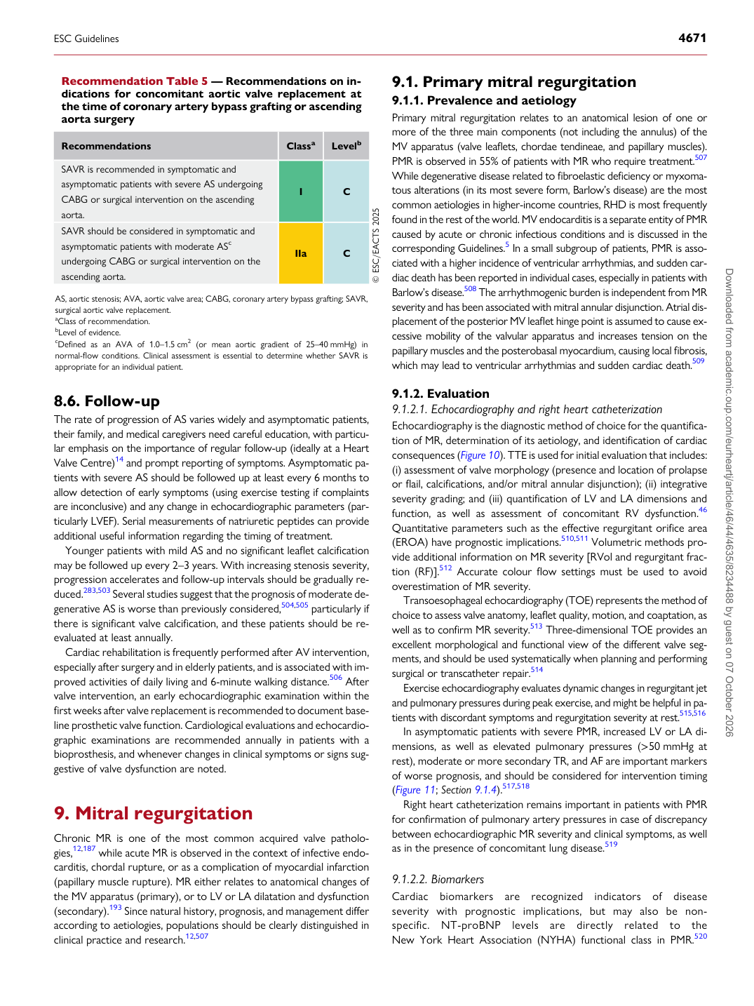
Recommendation Table 5 — Recommendations on indications for concomitant aortic valve replacement at the time of coronary artery bypass grafting or ascending aorta surgery

Recommendations Classa Levelb

SAVR is recommended in symptomatic and asymptomatic patients with severe AS undergoing CABG or surgical intervention on the ascending aorta. I C SAVR should be considered in symptomatic and asymptomatic patients with moderate ASc undergoing CABG or surgical intervention on the ascending aorta. IIa C © ESC/EACTS 2025

AS, aortic stenosis; AVA, aortic valve area; CABG, coronary artery bypass grafting; SAVR, surgical aortic valve replacement. aClass of recommendation. bLevel of evidence. cDefined as an AVA of 1.0–1.5 cm2 (or mean aortic gradient of 25–40 mmHg) in normal-flow conditions. Clinical assessment is essential to determine whether SAVR is appropriate for an individual patient.

ESC Guidelines                                                                                                                                                                                          4671

Downloaded from academic.oup.com/eurheartj/article/46/44/4635/8234488 by guest on 07 October 2026