p.30 抽出テキスト
Patient with suspected AS

Vmax ≥4 m/s and mean PG ≥40 mmHg

AVA ≤1 cm2 (AVAi ≤0.6 cm2/m2) AVA ≤1 cm2 (AVAi ≤0.6 cm2/m2)

Non-severe AS

Exclude measurement errorsa

SVi ≤35 mL/m2 Reversible high flowb

LVEF ≥50%

AVCS >1200 AU (female) >2000 AU (male)

Severe AS Severe AS

DSE with flow reserved

and AVA >1 cm2

Integrated assessmentf Pseudo-severe AS

Normal flow low-gradient AS (severe AS less likely)c Reassess under

normal flow

DSE with flow reserved and AVA ≤1 cm2 and/ore AVCS >1200 AU (female) >2000 AU (male)

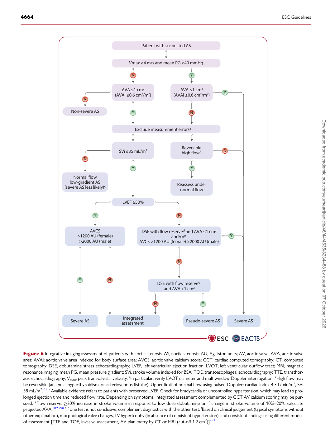
Figure 6 Integrative imaging assessment of patients with aortic stenosis. AS, aortic stenosis; AU, Agatston units; AV, aortic valve; AVA, aortic valve area; AVAi; aortic valve area indexed for body surface area; AVCS, aortic valve calcium score; CCT, cardiac computed tomography; CT, computed tomography; DSE, dobutamine stress echocardiography; LVEF, left ventricular ejection fraction; LVOT, left ventricular outflow tract; MRI, magnetic resonance imaging; mean PG, mean pressure gradient; SVi, stroke volume indexed for BSA; TOE, transoesophageal echocardiography; TTE, transthoracic echocardiography; Vmax, peak transvalvular velocity. aIn particular, verify LVOT diameter and multiwindow Doppler interrogation. bHigh flow may be reversible (anaemia, hyperthyroidism, or arteriovenous fistulae). Upper limit of normal flow using pulsed Doppler: cardiac index 4.3 L/min/m2, SVi 58 mL/m2.288 cAvailable evidence refers to patients with preserved LVEF. Check for bradycardia or uncontrolled hypertension, which may lead to prolonged ejection time and reduced flow rate. Depending on symptoms, integrated assessment complemented by CCT AV calcium scoring may be pursued. dFlow reserve: ≥20% increase in stroke volume in response to low-dose dobutamine or if change in stroke volume of 10%–20%, calculate projected AVA.289,290 eIf one test is not conclusive, complement diagnostics with the other test. fBased on clinical judgement (typical symptoms without other explanation), morphological valve changes, LV hypertrophy (in absence of coexistent hypertension), and consistent findings using different modes of assessment [TTE and TOE, invasive assessment, AV planimetry by CT or MRI (cut-off 1.2 cm2)]291. Downloaded from academic.oup.com/eurheartj/article/46/44/4635/8234488 by guest on 07 October 2026