p.64 抽出テキスト
11.9.4 Therapy

Medical therapy is considered for specific aetiologies, such as TB, to prevent progression to constriction.558 Antituberculosis drugs may significantly reduce the risk of constriction from >80% to <10%.569 Anti-inflammatory therapy may help to resolve transient constriction in 10%–20% of cases of AP.121,122 In such cases, to identify patients with potentially reversible forms of constriction, pericarditis should be excluded by testing markers of inflammation (e.g. C-reactive protein) or looking for imaging evidence of pericardial inflammation (e.g. pericardial oedema and/or LGE on CMR). In advanced cases, medical therapy is supportive of controlling symptoms of congestion, but surgery should never be delayed to improve outcomes. Pericardiectomy is the standard therapy for symptomatic chronic constriction without inflammation or not responding to antiinflammatory therapy (see Section 6.4.2). For milder disease or more advanced disease, or for those with radiation-induced disease, reduced EF, or advanced renal disease, caution is necessary. Radical pericardiectomy rather than anterior phrenic-to-phrenic pericardiectomy is warranted for the best outcomes.311,635–637 In general, pericardiectomy has been seen as a high-risk surgery with operative mortality of 6%–10%. However, recent studies show better results for early surgery in high-volume centres. The risk is related to the aetiologies and other comorbidities. Idiopathic causes have the lowest risk of <1.5%.638–640 11.10 Effusive–constrictive pericarditis

11.10.1. Introduction In ECP, pericardial fluid is present, and the scarred pericardium puts pericardial fluid under increased pressure, constricts the cardiac volumes, and leads to signs of CTP.116,117 Classically it is defined haemodynamically as failure of the RAP to fall by 50% or to a level below 10 mmHg after pericardiocentesis.117 Modern multimodality imaging allows a non-invasive diagnosis without cardiac catheterization.

11.10.2 Aetiology

Limited data have been published on ECP, and it appears to be a common condition in developing countries.117 Idiopathic cases are most commonly found in developed countries, while TB appears to be the most common cause in developing countries. The other reported causes are neoplasia, radiation, chemotherapy, infections (mainly TB and purulent type), and post-surgical pericardial disease.641

11.10.3 Presentation

Clinical features of either CP or CTP, or sometimes both, are typically present in patients with ECP. In patients who appear to have uncomplicated CTP, the diagnosis usually becomes evident during pericardiocentesis.117 Tricuspid regurgitation or right HF may cause persistently elevated RAP after efficient pericardiocentesis.

11.10.4 Diagnosis

To diagnose ECP, non-invasive imaging may be valuable.641 Thickening of the epicardial–pericardial layer is responsible for the constrictive component. After pericardiocentesis for CTP, careful detection of constriction on Doppler findings can be reported.124 Without haemodynamic monitoring, ECP can also be suspected on echocardiography and CMR (see Supplementary data online, Table S14). Cardiovascular magnetic resonance may provide helpful information, especially in differentiating constriction from RCM (Table S14). An accurate evaluation of ventricular interdependence and septal bounce can be made by assessing ventricular coupling with echocardiography and/or real-time cine CMR.115 Echo-Doppler findings indicating effusive constriction may be detected prior to pericardiocentesis.116,124 Currently, the diagnosis can be made using multimodality imaging without cardiac catheterization.

11.10.5 Treatment

Effusive constriction can often resolve spontaneously or be treated with anti-inflammatory therapies in the case of evidence of transient constriction.641 In resistant cases, visceral pericardiectomy must be performed, since the epicardial layer is responsible for constricting the heart. The visceral component of pericardiectomy requires sharp dissection of many small fragments until ventricular motion is improved. Only pericardial centres with experience and competency should perform pericardiectomy for CP.

12 Age- and sex-related aspects in

inflammatory myopericardial syndrome

12.1 Sex distribution

Acute myocarditis is more prevalent in males (75%–84%).17,28,73,78,642 On the contrary, the male-to-female ratio is similar in fulminant cases (46% females).56,75,643 Sex differences in pericarditis are less evident,

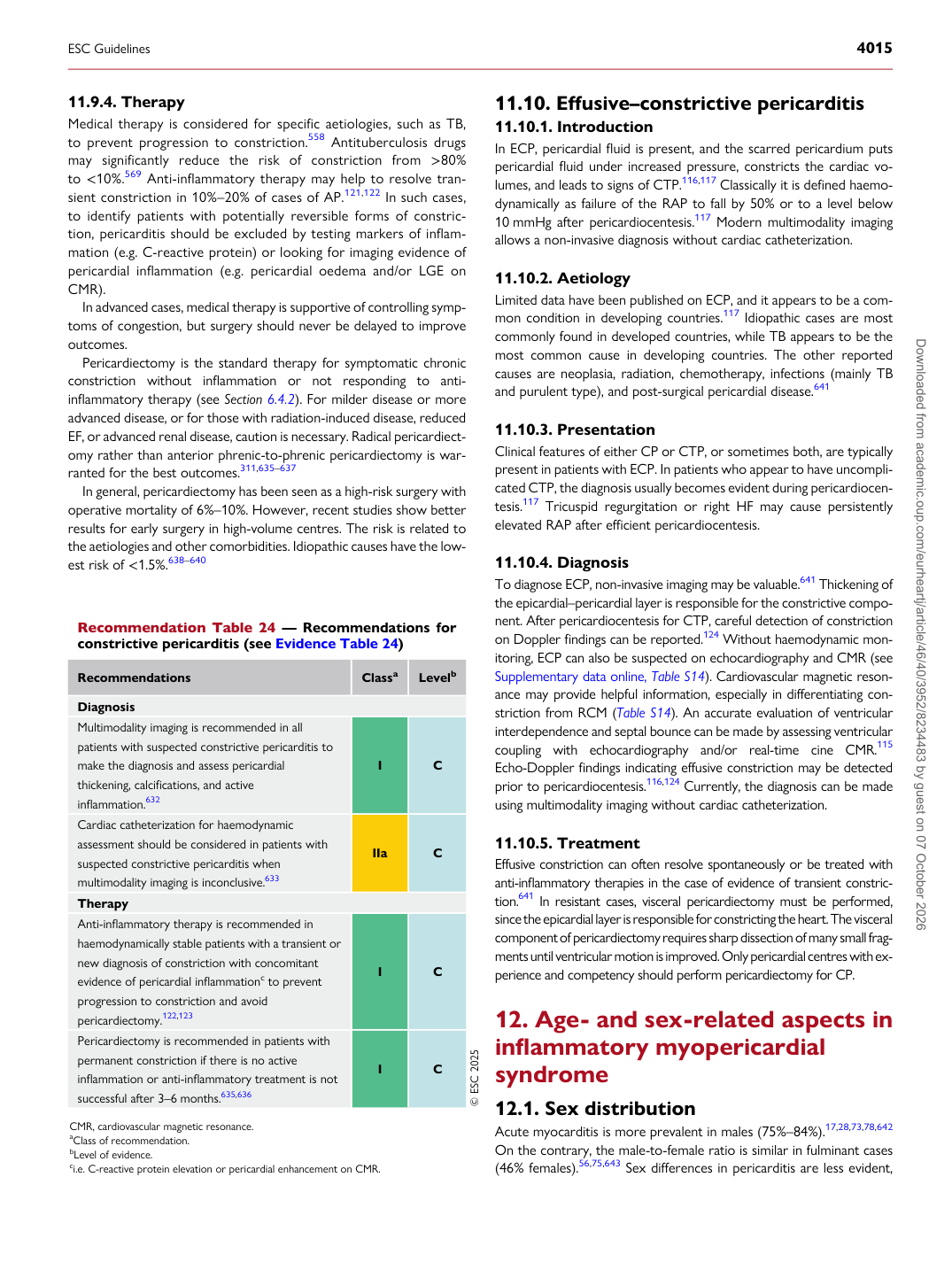
Recommendation Table 24 — Recommendations for constrictive pericarditis (see Evidence Table 24)

Recommendations Classa Levelb

Diagnosis Multimodality imaging is recommended in all patients with suspected constrictive pericarditis to make the diagnosis and assess pericardial thickening, calcifications, and active inflammation.632 I C Cardiac catheterization for haemodynamic assessment should be considered in patients with suspected constrictive pericarditis when multimodality imaging is inconclusive.633 IIa C Therapy Anti-inflammatory therapy is recommended in haemodynamically stable patients with a transient or new diagnosis of constriction with concomitant evidence of pericardial inflammationc to prevent progression to constriction and avoid pericardiectomy.122,123 I C Pericardiectomy is recommended in patients with permanent constriction if there is no active inflammation or anti-inflammatory treatment is not successful after 3–6 months.635,636 I C © ESC 2025

CMR, cardiovascular magnetic resonance. aClass of recommendation. bLevel of evidence. ci.e. C-reactive protein elevation or pericardial enhancement on CMR.

ESC Guidelines                                                                                                                                                                                          4015

Downloaded from academic.oup.com/eurheartj/article/46/40/3952/8234483 by guest on 07 October 2026