p.63 抽出テキスト
The diagnosis of CTP identifies high-risk patients with an increased risk of complications during follow-up and a high probability of a nonviral aetiology. The patients should be admitted for therapy and monitoring.1,105 The definitive therapy is pericardiocentesis, which should be performed urgently according to the clinical presentation.

11.8.3 Therapy

The treatment for CTP is drainage of the pericardial fluid, preferably by needle pericardiocentesis with the use of echocardiographic or fluoroscopic guidance, and should be performed without delay in unstable patients. Alternatively, drainage is performed by a surgical approach, especially in situations such as purulent pericarditis, or urgent situations with bleeding into the pericardium. Pericardiocentesis should be performed by experienced operators trained/certified in acute cardiovascular care and carries a variable risk of complications from 4% to 10%, depending on type of monitoring, operator’s skill, and setting (emergent vs urgent vs elective). The most common complications include arrhythmias, coronary artery or cardiac chamber puncture, haemothorax, pneumothorax, pneumopericardium, and hepatic injury.1 Mechanical ventilation with positive airway pressure should be avoided in CTP. In addition, diuretic therapy should be avoided, and temporary fluid administration can be helpful to stabilize the patient while waiting for urgent pericardiocentesis.

11.8.4 Outcomes and prognosis

The prognosis of CTP is essentially related to its aetiology. Cardiac tamponade in patients with cancer and metastatic involvement of the pericardium has a bad short-term prognosis, since this is a sign of advanced disease. In contrast, patients with CTP and a final diagnosis of idiopathic pericarditis generally have a good long-term prognosis.1,105

11.9 Pericardial constriction and

constrictive pericarditis (calcified and non-calcified) 11.9.1. Introduction Pericardial constriction is a chronic condition usually characterized by a thickened, fibrotic, and often calcified pericardium that leads to impaired diastolic filling of the heart. Constrictive pericarditis is constriction associated with pericarditis.114,119,347 There is a high risk of progression in bacterial pericarditis (20%–30%), especially purulent pericarditis, intermediate risk in immune-mediated pericarditis and neoplastic pericardial diseases (2%–5%), and a low risk in viral and idiopathic pericarditis (<1%).347 Cases of CP after pericardiectomy have been reported, especially after partial pericardiectomy.627–630 In developed countries, the most common reported causes are: viral or idiopathic (42%–49%); post-cardiac surgery (11%–37%); post-radiation therapy, mostly for breast cancer or Hodgkin’s disease (9%–31%); connective tissue disorders (3%–7%); post-infectious causes (purulent pericarditis or TB in 3%–6%); and miscellaneous causes (<10%). TB is a major cause of CP in developing countries.630,631

11.9.2 Presentation

The classical clinical presentation comprises signs and symptoms of right HF with preserved biventricular function without previous or concomitant myocardial disease. Subacute/chronic inflammatory pericarditis can evolve into CP, and the duration of progression from initial pericardial inflammation and the onset of constriction is variable.347 Systolic dysfunction due to myocardial fibrosis or atrophy can aggravate the haemodynamic impairment in more advanced cases. In up to 20% of cases, constriction might be present with normal pericardial thickness, while advanced and classic cases demonstrate prominent pericardial thickening and calcifications.119

11.9.3 Diagnosis

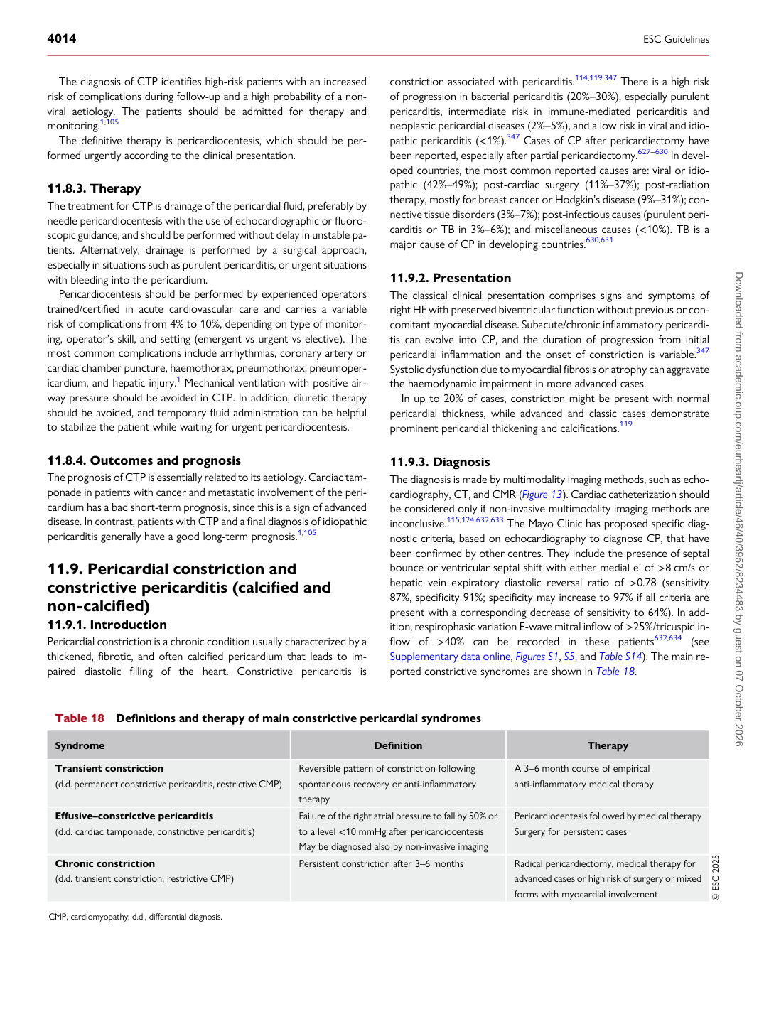
The diagnosis is made by multimodality imaging methods, such as echocardiography, CT, and CMR (Figure 13). Cardiac catheterization should be considered only if non-invasive multimodality imaging methods are inconclusive.115,124,632,633 The Mayo Clinic has proposed specific diagnostic criteria, based on echocardiography to diagnose CP, that have been confirmed by other centres. They include the presence of septal bounce or ventricular septal shift with either medial e’ of >8 cm/s or hepatic vein expiratory diastolic reversal ratio of >0.78 (sensitivity 87%, specificity 91%; specificity may increase to 97% if all criteria are present with a corresponding decrease of sensitivity to 64%). In addition, respirophasic variation E-wave mitral inflow of >25%/tricuspid inflow of >40% can be recorded in these patients632,634 (see Supplementary data online, Figures S1, S5, and Table S14). The main reported constrictive syndromes are shown in Table 18.

Table 18 Definitions and therapy of main constrictive pericardial syndromes

Syndrome Definition Therapy

Transient constriction (d.d. permanent constrictive pericarditis, restrictive CMP) Reversible pattern of constriction following spontaneous recovery or anti-inflammatory

therapy A 3–6 month course of empirical anti-inflammatory medical therapy

Effusive–constrictive pericarditis (d.d. cardiac tamponade, constrictive pericarditis) Failure of the right atrial pressure to fall by 50% or

to a level <10 mmHg after pericardiocentesis May be diagnosed also by non-invasive imaging Pericardiocentesis followed by medical therapy

Surgery for persistent cases

Chronic constriction (d.d. transient constriction, restrictive CMP) Persistent constriction after 3–6 months Radical pericardiectomy, medical therapy for

advanced cases or high risk of surgery or mixed forms with myocardial involvement © ESC 2025

CMP, cardiomyopathy; d.d., differential diagnosis. Downloaded from academic.oup.com/eurheartj/article/46/40/3952/8234483 by guest on 07 October 2026