p.61 抽出テキスト
Additional symptoms due to the compression of adjacent structures include nausea (diaphragm), dysphagia (oesophagus), hoarseness (recurrent laryngeal nerve), and hiccups (phrenic nerve).118,619

11.7.3 Diagnosis

Physical examination may be normal in patients without haemodynamic compromise. Echocardiography is the primary diagnostic tool for diagnosis, sizing, assessment of haemodynamic effects, and follow-up (see Supplementary data online, Figure S5).115,124 Advanced imaging (CT and CMR) provides valuable information concerning local inflammation, masses, associated chest abnormalities (e.g. pectus excavatum), loculated effusions, and systemic diseases.196,198,620 Pericardial effusion is often associated with known or unknown medical conditions (e.g. hypothyroidism) in up to 60% of cases, especially if moderate/large.615

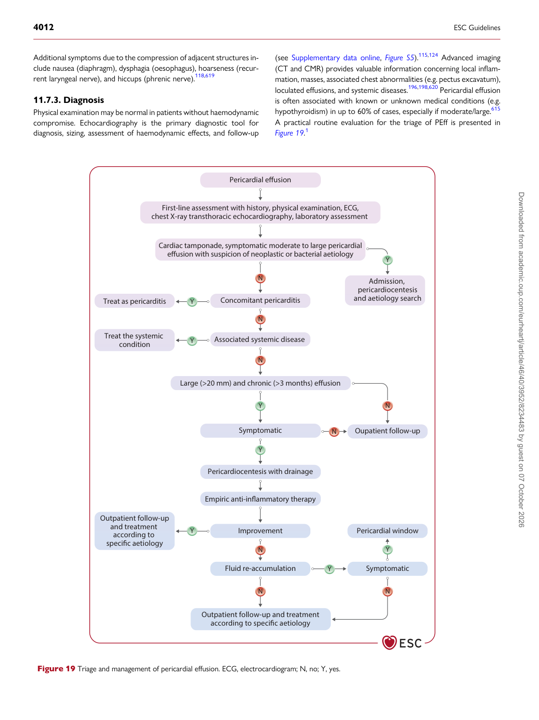
A practical routine evaluation for the triage of PEff is presented in Figure 19.1

Concomitant pericarditis Treat as pericarditis

Associated systemic disease

Pericardiocentesis with drainage

Empiric anti-inflammatory therapy

Improvement

Outpatient follow-up

and treatment according to specific aetiology Pericardial effusion

First-line assessment with history, physical examination, ECG, chest X-ray transthoracic echocardiography, laboratory assessment

Cardiac tamponade, symptomatic moderate to large pericardial

effusion with suspicion of neoplastic or bacterial aetiology

Admission, pericardiocentesis and aetiology search

Large (>20 mm) and chronic (>3 months) effusion

Pericardial window

Treat the systemic

condition

Symptomatic Oupatient follow-up

Symptomatic Fluid re-accumulation

Outpatient follow-up and treatment

according to specific aetiology

Figure 19 Triage and management of pericardial effusion. ECG, electrocardiogram; N, no; Y, yes. Downloaded from academic.oup.com/eurheartj/article/46/40/3952/8234483 by guest on 07 October 2026