p.60 抽出テキスト
the predominant associated lesions are empyema (50%) or pneumonia (33%). In immunosuppressed patients or following thoracic surgery, the most frequently encountered bacterial/fungal agents are Staphylococcus aureus (30%) and fungi (20%), while anaerobic bacteria, originating from the oropharynx, have also been reported.607,608 It should be stressed that in the modern era of iatrogenic and HIV-associated immunosuppression, more unusual organisms may account for pericardial infection.1,607

11.5.1 Presentation

Purulent pericarditis has generally manifested as a serious febrile disease, and the underlying sepsis may predominate, including septic shock.606–608 Constrictive pericarditis complicates 20%–30% of cases.347

11.5.2 Diagnosis

Suspicion of purulent pericarditis is an indication for pericardial drainage independently from the haemodynamic status. Surgical pericardial drainage is recommended to allow complete removal of the fluid and prevent its organization and progression to constriction. Low pericardial-to-serum glucose ratio (mean 0.3) and elevated pericardial fluid white cell count with a high proportion of neutrophils (mean cell count 2.8/mL, 92% neutrophils) differentiate purulent from tuberculous (glucose ratio 0.7, mean cell count 1.7/mL, 50% neutrophils) and neoplastic pericarditis (glucose ratio 0.8, mean cell count 3.3/mL, 55% neutrophils). Fluid must be sent for bacterial, fungal, and tuberculous studies along with blood and tissue samples, as guided by the clinical presentation.1

11.5.3 Therapy

Intravenous antimicrobial therapy should be started empirically until microbiological results are available. Afterwards, pathogen-directed antibiotic therapy is warranted based on antibiotic susceptibility.1 The duration of antibiotic therapy should be individualized until fever and clinical signs resolve, with the minimum period of administration being 3 weeks. Purulent effusions are often heavily loculated and likely to rapidly reaccumulate. Subxiphoid pericardiotomy and rinsing of the pericardial cavity should be considered.15 This allows more complete drainage of the effusion, since loculations can be manually lysed. Intrapericardial fibrinolysis is a possible treatment for cases of loculated effusions to achieve adequate drainage.609 Fibrinolysis has been shown to prevent complications in approximately 86% of cases without fatal complications.573,609,610 In the unfortunate case that percutaneous measures fail to control infection, and worsening HF occurs along with constrictive physiology features, referral to surgical pericardiectomy is advised.610 11.6 Incessant and recurrent pericarditis

The TF proposes to adopt the term ‘incessant’ for cases of persistent symptoms without clear remission, and thus also to include cases of symptom persistence/recurrence during drug tapering. Recognition of these cases is of paramount importance, since they can directly progress to CP within a few months.104 Recurrent pericarditis is defined as a relapse after a documented first episode of AP, a symptom-free interval, complete discontinuation of anti-inflammatory therapy, and evidence of subsequent recurrence of pericarditis. Recurrence usually occurs within 18 months from the index episode, and is the most common and problematic complication of AP, ranging from 15% to 30% of cases after a first episode of pericarditis. It may increase to 50% after a first recurrence in patients with inadequate treatment of the first episode (e.g. too fast tapering, not treated with colchicine, treated with corticosteroids).25,611 In developed countries, the aetiology of recurrences is poorly known, and most cases are labelled as idiopathic. Historically, most cases were probably related to an autoinflammatory/autoimmune pathogenesis. This is based on the inflammatory/non-inflammatory phenotype, the latency from the acute attack (several weeks after the index episode), the presence of autoantibodies, and the response to anti-inflammatory/ immunosuppressive treatments.52,283,284,287

11.7 Inflammatory and non-inflammatory

pericardial effusion The normal pericardial sac contains 10–50 mL of pericardial fluid, a plasma ultrafiltrate that acts as a lubricant between the pericardial layers. Any pathological process that causes pericardial inflammation increases the amount of fluid, causing PEff (exudate). Alternative mechanisms causing PEff include decreased reabsorption following increased systemic venous pressure, as observed in congestive HF or pulmonary hypertension (transudate) or alternatively, decreased lymphatic drainage.612 The regimen of pressures in the epicardium and pericardial cavity is shown in Supplementary data online, Figure S4.

11.7.1 Classification and aetiology

The reported incidence and prevalence of PEff in developed countries is estimated at 3% and 6%–9%, respectively, based on echocardiographic laboratory records.613 Pericardial effusion was detected in 14% of subjects in a prospective registry of consecutive patients who underwent chest CT.198 The classification of PEff is shown in Table 10. Echocardiography provides semiquantitative assessment of the effusion size, which is useful for estimating the risk of having a non-idiopathic aetiology and complications (see Supplementary data online, Figure S5).105 In the past three decades, six major surveys have been published on the characteristics of moderate to large PEff (see Supplementary data online, Table S13).556,614–618 According to these series, many cases remain idiopathic in developed countries (up to 50%), while additional aetiologies include cancer (10%–32%), infections (15%–30%), iatrogenic causes (15%–20%), and connective tissue diseases (5%–15%), whereas TB is the dominant cause in developing countries (>60%), where TB is endemic.558

11.7.2 Presentation

A significant proportion of patients with PEff are asymptomatic, and PEff is an incidental finding during routine examination. The clinical presentation of PEff varies according to the speed of accumulation of fluid (see Supplementary data online, Figure S4). Classic symptoms include exertional dyspnoea progressing to orthopnoea, chest pain, and feeling of fullness.

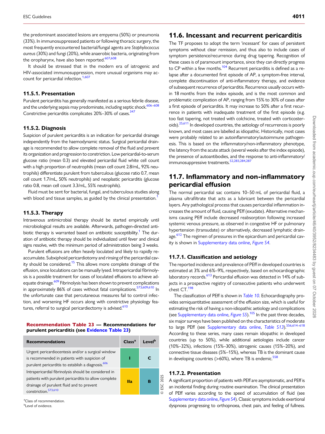
Recommendation Table 23 — Recommendations for purulent pericarditis (see Evidence Table 23)

Recommendations Classa Levelb

Urgent pericardiocentesis and/or a surgical window is recommended in patients with suspicion of purulent pericarditis to establish a diagnosis.606 I C Intrapericardial fibrinolysis should be considered in patients with purulent pericarditis to allow complete drainage of purulent fluid and to prevent constriction.573,610 IIa B © ESC 2025 aClass of recommendation. bLevel of evidence.

ESC Guidelines                                                                                                                                                                                          4011

Downloaded from academic.oup.com/eurheartj/article/46/40/3952/8234483 by guest on 07 October 2026