p.56 抽出テキスト
also important for the pathogenesis of inflammatory CMP.9 However, the rates, risk factors, and other characteristics of cases progressing from AM to chronic myocarditis or inflammatory CMP remain unknown.

10.1 Presentation

Patients with inflammatory CMP may present with new-onset/acute, subacute, or chronic HF. Although myocarditis resolves in >50% of cases, even spontaneously, approximately 25% of cases may present with persistent ventricular dysfunction, and more severe cases may progress to DCM.10

10.2 Diagnosis

A variety of clinical assessment, ECG, multimodality imaging, and, if needed, EMB can be used to reach a diagnosis. Persistent or chronic myocardial inflammation should be suspected in cases with nonischaemic ventricular dysfunction associated with low QRS voltages, and persistent mild elevation of hs-TnT/TnI. The absence of significant remodelling may be observed in the early phase of the disease. This suspicion can be confirmed by multimodality imaging (echocardiography and CMR) and EMB, especially in the presence of myocardial oedema observed at CMR. Inflammatory CMP may be familial in rare cases. In these patients, genetic testing should be discussed.

10.3 Therapy

Guideline HF treatment is recommended in patients with inflammatory CMP.12,259 Medical therapy should be tailored to the underlying cause in case of systemic disorders, especially in autoimmune diseases. Only one RCT showed an improvement of cardiac function with immunosuppressive treatment in patients with virus-negative inflammatory CMP.551 Other non-randomized studies, often derived from singlecentre and/or retrospective studies,34,131,551–555 suggest the efficacy of immunosuppression on top of optimized HF therapy, and support its safety. In patients with different immune-mediated myocarditis and HF, immunosuppressive therapy was safe.34,131,552–555 Therefore, the treatment should be based on histological/immunohistological and viral PCR examination of the biopsy specimens. In untreated cases, progression to DCM and transplantation or death are observed.10,39 11 Specific types of pericarditis

11.1 Tuberculous pericarditis

In high-income countries, TB pericarditis accounts for up to 4% of pericardial disease, but it is increasing due to immigration. On the other hand, TB is the cause of clinically significant PEff in approximately 90% of HIV-infected and 50%–70% of non-HIV-infected individuals living in developing countries with a high prevalence of TB.556 It should be emphasized that most of the information on TB pericarditis comes from endemic areas in low-income countries and immunosuppressed patients.1 Notably, the disease can occur at any age, and men are affected more frequently than women.48 Tuberculous pericarditis has an overall mortality rate of 17%–40% at 6 months after diagnosis.557

11.1.1 Presentation

Clinical presentations of TB pericardial involvement include AP (most commonly with PEff), ECP, and CP, with these manifestations representing consecutive stages of a unique process rather than independent conditions.558,559 Specifically, the evolution of TB pericarditis encompasses four stages (Figure 18). The first stage or dry stage of AP is without effusion, the second stage with PEff (this is the form most commonly encountered in clinical practice) in up to 80% of cases, the third stage with progressive absorption of effusion, and the fourth stage with pericardial constriction. In the effusive phase, pericardiocentesis usually yields bloody effusion, and effusive–constrictive physiology may emerge after pericardial drainage. At this stage, lymphocytic exudate is mainly observed.560 The duration of each stage varies and depends on individual factors (e.g. HIV status), and early treatment may prevent progression.

11.1.2 Diagnosis

The diagnosis of TB pericarditis is challenging and in 15%–20% of cases the diagnosis may be missed.561 A ‘definite’ diagnosis of TB is based on the presence of tubercle bacilli in the pericardial fluid or on a histological sample of the pericardium, by culture or by PCR (Xpert MTB/RIF and RIF ultra) testing. A ‘probable’ diagnosis is made when there is proof of TB elsewhere in a patient with unexplained pericarditis, a lymphocytic pericardial exudate with elevated unstimulated IFN-γ (uIFN-γ), adenosine deaminase or lysozyme levels, and/or an appropriate response to antituberculosis therapy in endemic areas.558 In a meta-analysis, the sensitivity and specificity of adenosine deaminase, which is probably the most widely used biomarker, were 90% and 86%, respectively, for the diagnosis of TB pericarditis.562 Xpert MTB/RIF is highly specific (100%); however, it has a sensitivity of 64%, which increases to 78% when pericardial biopsy samples are employed.563,564 Pericardial fluid culture shows low sensitivity (53%–75%) and it is also time-consuming, needing more than 3 weeks to yield results. Interferon gamma offers superior accuracy for the diagnosis of microbiologically confirmed TB pericarditis compared with the adenosine deaminase (ADA) assay and the Xpert MTB/RIF test.563 In a meta-analysis, values of uIFN-γ in the pericardial fluid from studies using thresholds of 14–200 pg/mL showed very high sensitivity (97%) and specificity (99%) for the diagnosis of TB pericarditis, with a positive likelihood ratio of 187 and a negative likelihood ratio of 0.03.565 On the other hand, the tuberculin skin test and IFN-γ release assays are of limited value, especially in areas with a high burden of TB, since they cannot discriminate between previous exposure to Mycobacterium and active disease.566 A detailed approach is outlined in Supplementary data online, Table S12.

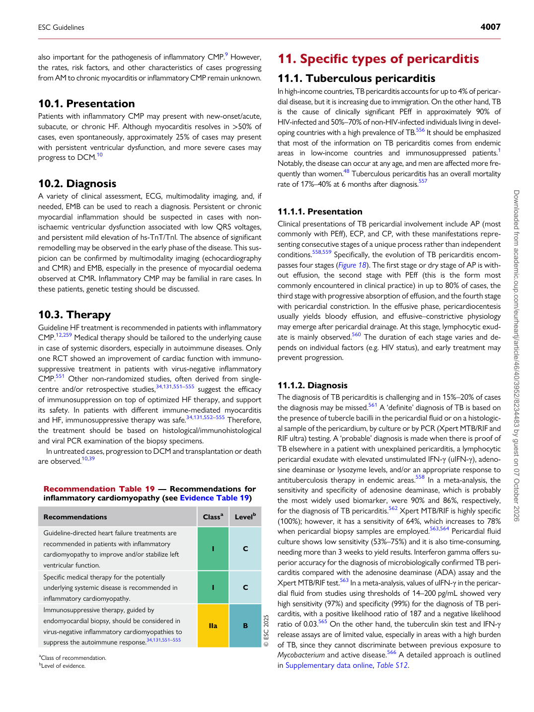
Recommendation Table 19 — Recommendations for inflammatory cardiomyopathy (see Evidence Table 19)

Recommendations Classa Levelb

Guideline-directed heart failure treatments are recommended in patients with inflammatory cardiomyopathy to improve and/or stabilize left ventricular function. I C Specific medical therapy for the potentially underlying systemic disease is recommended in inflammatory cardiomyopathy. I C Immunosuppressive therapy, guided by endomyocardial biopsy, should be considered in virus-negative inflammatory cardiomyopathies to suppress the autoimmune response.34,131,551–555 IIa B © ESC 2025 aClass of recommendation. bLevel of evidence.

ESC Guidelines                                                                                                                                                                                          4007

Downloaded from academic.oup.com/eurheartj/article/46/40/3952/8234483 by guest on 07 October 2026