p.53 抽出テキスト
9.5 Specific infectious myocarditis

(Viruses, Lyme, Chagas) 9.5.1. Viruses A large variety of infectious agents (Supplementary data online, Table S1) can cause myocardial inflammation, but viruses are considered the most common cause among infectious forms of myocarditis. Data on the true prevalence of viral myocarditis are difficult to determine, since EMB is performed only in a subgroup of high-risk patients and in complicated cases. Moreover, a positive viral serological test does not imply myocardial infection and is not routinely recommended. Exceptions are HIV and HCV and for specific infections such as Lyme disease.10 The epidemiology of viral myocarditis has changed during the past three decades; while enteroviruses and adenoviruses have been considered the most common causes of viral myocarditis,468 vasculotropic viruses such as B19V,63,66,468 and lymphotropic viruses such as HHV-6 are increasingly found in EMB samples.342,469 The prognostic relevance of persistent B19V infection with low viral copy numbers remains controversial because its presence in myocardial tissue may not have direct pathogenetic significance,470,471 in contrast to acute B19V infection.228

9.5.1.1 Presentation

Symptoms vary according to the underlying cause and severity of disease. However, it generally follows the presentation of acute and chronic stages, as presented in Section 4.

9.5.1.2 Diagnosis

The diagnostic approach should follow the description outlined in Section 4 (Figures 5 and 6). The presence of more than 500 copies/μg of DNA in EMB is considered as the threshold for virus-related myocardial inflammation, and usually guides therapeutic choices (e.g. starting immunosuppression). Viral PCR analysis in EMB samples is performed to better characterize the pathological substrate causing myocarditis. If immunosuppression is needed, viral presence has to be ruled out by EMB.10 Contemporary PCR analysis for viral presence in EMB specimens and blood samples is utilized to rule out possible tissue contamination.10,469

9.5.1.3 Therapy

Data are insufficient to support antiviral therapy for AM, as well as treatment with IVIG for presumed viral myocarditis.472 Anti-herpesvirus drugs in patients with CMV or HHV-6 infection can be considered, although their efficacy has not been directly studied in patients with myocarditis.10 Treatment with type I IFN in patients with enterovirus has been suggested to increase viral clearance, but large trials are missing.273,473–475 Antiviral therapies have to be prescribed and managed in collaboration with an infectious diseases expert with shared decisionmaking in the IMPS team.1,472

9.5.1.3.1 Influenza virus. Supportive management is the backbone of

severe influenza treatment. Myocardial involvement is rare, but should be considered in patients of any age with chest pain, tachycardia, and haemodynamic instability within 2–4 weeks from symptom onset. The effect of antiviral therapy on myocarditis is not clear, although oseltamivir is utilized in both inpatient and outpatient settings. Peramivir and zanamivir are used only for hospitalized patients. There are individual case reports of successful treatment with neuraminidase inhibitors.476–479 Peramivir has often been used in combination with either oseltamivir480,481 or zanamivir.482

9.5.1.3.2 Human immunodeficiency virus. The pathogenesis of

HIV-associated myocarditis involves direct viral infection, co-infection with other viruses or bacteria, cytokine activity, side effects of highly active antiretroviral therapy (HAART), immune system dysregulation, and/or ischaemia. If HIV-associated myocarditis is diagnosed, therapy for HIV infection has to be started or continued. Highly active antiretroviral therapy has been reported to significantly decrease the incidence of cardiac involvement, especially pericarditis, arrhythmias, and DCM compared with patients treated with nucleoside reverse transcriptase inhibitors in a retrospective study of 1042 patients.483

9.5.2 Lyme carditis

Lyme carditis may be an early manifestation of Lyme borreliosis, a tickborne disease, caused by Borrelia spp., which is one of the most common vector-borne diseases in the northern hemisphere.

9.5.2.1 Presentation

In cases of early disseminated infection, AM with possible associated pericarditis may occur, but LC is typically characterized by varying degrees of AVB, with third-degree AVB in approximately 25% of cases.484 Chronic LC often shows manifested persistent symptoms and signs, such as fatigue, cognitive dysfunction, headaches, sleep disturbance, neuropsychiatric and musculoskeletal symptoms, electrical conduction delays, and DCM.

9.5.2.2 Diagnosis

The suspicious index in LC score has been proposed to evaluate the likelihood of high-degree AVB to be caused by LC. It allows categorization of patients into low-, intermediate- and high-risk categories.485 Continuous ECG monitoring is required for patients presenting with syncope or with a PR interval of >300 ms, since this is a high-risk marker of progression to complete AVB.484 Serological tests are recommended for patients presenting with high-degree AVB and suspected LC.

9.5.2.3 Therapy

Atrioventricular block is often transient and typically resolves within 1–2 weeks of antibiotic treatment, therefore permanent pacemaker placement is not warranted for AVB associated with LC before starting therapy. Intravenous therapy with ceftriaxone is the recommended first-line treatment for patients with LC and AV disturbances,486 followed by oral antibiotics (doxycycline, amoxicillin, or cefuroxime) for

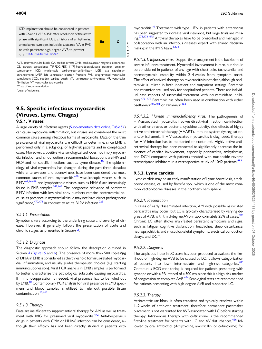
ICD implantation should be considered in patients with CS and LVEF >35% after resolution of the active phase with significant LGE, a history of arrhythmias, unexplained syncope, inducible sustained VA at PVS, or with persistent high-degree AVB to prevent SCD.416,434,452,453,456–464,467 IIa C © ESC 2025

AVB, atrioventricular block; CA, cardiac arrest; CMR, cardiovascular magnetic resonance; CS, cardiac sarcoidosis; 18F-FDG-PET, [18F]-fluorodeoxyglucose positron emission tomography; ICD, implantable cardioverter-defibrillator; LGE, late gadolinium enhancement; LVEF, left ventricular ejection fraction; PVS, programmed ventricular stimulation; SCD, sudden cardiac death; VA, ventricular arrhythmias; VF, ventricular fibrillation; VT, ventricular tachycardia. aClass of recommendation. bLevel of evidence.

Downloaded from academic.oup.com/eurheartj/article/46/40/3952/8234483 by guest on 07 October 2026