p.52 抽出テキスト
9.4 Myocarditis in sarcoidosis

Cardiac sarcoidosis is a form of inflammatory heart disease, coexisting with systemic sarcoidosis in 5%–10% of cases,406 or rarely, in isolation. The aetiology of sarcoidosis is not well understood, but may involve an autoimmune response after certain trigger(s) and genetic predisposition, which leads to the formation of non-caseating granulomas in multiple organs, typically in the lungs.407 On histopathology, the granuloma consists of CD68+ macrophages, CD68+ giant cells, CD3+ T lymphocytes, and CD20+ B lymphocytes. It is characterized by inflammation, oedema, lymphocytic infiltration, and replacement fibrosis.408 Cardiac sarcoidosis is often subclinical, with an estimated cardiac involvement in around 25% of cases on autopsy series in patients with sarcoidosis.409,410

9.4.1 Presentation

Manifestations of CS may range from cardiac inflammation, conduction system disease, frequent ectopy, tachyarrhythmia, and HF, to SCD.407 Cardiac sarcoidosis may mimic different cardiac phenotypes, including ARVC,254,411–413 DCM, and hypertrophic CMP (HCM), but may also occur in patients with normal cardiac function.414 Symptoms are nonspecific, and often related to arrhythmias, especially AVBs and VTs. The challenge is to identify patients at risk, since SCD due to malignant arrhythmia is common.415,416

9.4.2 Diagnosis

The diagnostic algorithm of CS is different in patients with or without known extracardiac sarcoidosis. Pathognomonic findings in noninvasive imaging are sufficient for the diagnosis of CS in cases with known systemic sarcoidosis.417 In contrast, the diagnostic pathway in primary CS is more challenging, but typical multimodality imaging patterns are usually sufficient. Cardiac sarcoidosis can manifest as abnormalities on ECG, echocardiography, CMR, PET, EMB, and cardiac and inflammatory blood biomarkers (see Section 5.4). Electrocardiogram or cardiac rhythm monitoring may demonstrate conduction system abnormalities (such as AVB or bundle-branch block) and arrhythmias, including VT.418 Echocardiography may reveal regional WMA in a non-coronary distribution, septal thinning, and ventricular aneurysms. Endomyocardial biopsy may be helpful in selected patients, but negative results do not rule out CS due to potential sampling errors.419 Endomyocardial biopsy guided by imaging or EAM may improve the diagnostic yield (see Section 5.12). Multimodality advanced cardiac imaging, such as CMR and FDG-PET, plays an increasing role in the management of suspected CS. Overall, compared with FDG-PET, CMR has higher sensitivity of 89%–95% vs 84%, respectively, but similar specificity of 78%–85% vs 82%, respectively, for the diagnosis of CS.408,420–428 On CMR, CS has a wide range of potential findings, and is often considered the ‘great mimicker’ of various cardiac conditions, but the interventricular septum is often involved408 and the ‘hook sign’429 is recognized to be associated with a high probability of CS. For more details, see Supplementary data online, Figure S3. A heavy burden of LGE is associated with a poorer prognosis and predisposes to VA, HF, and death in CS, and may guide clinical decision-making.430–436 Aside from LGE, the identification of ongoing myocardial inflammation is possible.30,437–441 Fluorodeoxyglucose-positron emission tomography also allows detection of cardiac inflammation in CS.426 It requires careful patient preparation to suppress physiological 18F-FDG uptake. The classical finding is focal 18F-FDG uptake in the basal septum, with a concordant perfusion defect. Perfusion defects in the active stage of CS may be due to microvascular obstruction by inflammatory oedema, necrosis, or both, and are not necessarily located in the same region as the 18F-FDG uptake.427

Hybrid PET-CMR may enable better detection of cardiac inflammation in a more comprehensive manner,440,442,443 but its availability is limited. Both CMR and PET may be used for imaging-guided EMB. The intensity and extent of FDG uptake on PET is of prognostic relevance.443–449 In a meta-analysis of 37 studies of patients with CS, LGE on CMR and FDG uptake on PET were both predictive of MACE (all-cause death, cardiac death and aborted SCD, VA, and HF hospitalization). Fluorodeoxyglucose-positron emission tomography has proved to be useful in predicting and monitoring the anti-inflammatory therapy response in sarcoidosis;204,450 however, the optimal timing for repeating PET studies for the assessment of treatment response is not well established. In addition to clinical assessment, imaging modalities should be used for the short- and long-term management of patients with CS.

9.4.3 Therapy

The treatment of CS is currently not standardized, and is based on patient-tailored assessment, often with multidisciplinary input.451 Treatment is targeted at controlling cardiac inflammation, myocardial scarring and dysfunction.13,431–436 First-line therapy includes corticosteroids (see Table 12). Since resolution of conduction abnormalities with anti-inflammatory treatment remains unpredictable, definitive cardiac pacing is usually needed. AVB and the need for cardiac pacing are associated with a higher risk of sustained VA, which reached 31% at 5 years in the Finnish registry.416,452,453 Thus, it appears reasonable to propose an ICD with pacing ability rather than a pacemaker in CS patients with pacing indications. The risk of VA during follow-up is high in observational studies, in patients with low LVEF,416,452,454,455 presence of LGE on CMR,434,456–459 and positive PVS.456,460–464 There is a lack of data on the relationship between scarring extent and arrhythmia risk. Positron emission tomography-computed tomography may also help to stratify patients.465 Heart transplantation could be considered in a subgroup of patients with CS, similarly to GCM.405

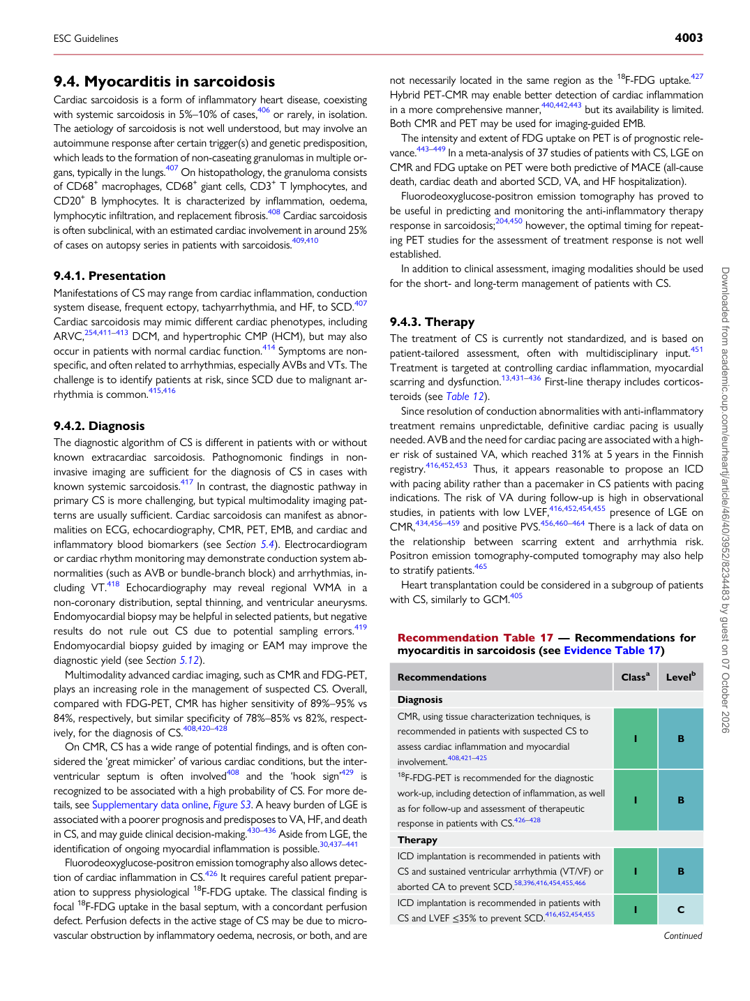
Recommendation Table 17 — Recommendations for myocarditis in sarcoidosis (see Evidence Table 17)

Recommendations Classa Levelb

Diagnosis CMR, using tissue characterization techniques, is recommended in patients with suspected CS to assess cardiac inflammation and myocardial involvement.408,421–425 I B 18F-FDG-PET is recommended for the diagnostic work-up, including detection of inflammation, as well as for follow-up and assessment of therapeutic response in patients with CS.426–428 I B Therapy ICD implantation is recommended in patients with CS and sustained ventricular arrhythmia (VT/VF) or aborted CA to prevent SCD.58,396,416,454,455,466 I B ICD implantation is recommended in patients with CS and LVEF ≤35% to prevent SCD.416,452,454,455 I C

Continued

ESC Guidelines                                                                                                                                                                                          4003

Downloaded from academic.oup.com/eurheartj/article/46/40/3952/8234483 by guest on 07 October 2026