p.47 抽出テキスト
7.2 Complications and outcomes for

pericarditis Risk stratification of patients with pericarditis must be performed at the initial presentation at the emergency department or in an ambulatory setting. Admission is recommended in high-risk cases.105,130 Non-high-risk cases can be managed as outpatients with a close followup within 1 to 2 weeks (Figure 7). ECG changes (mainly widespread ST-segment elevation) and troponin elevation were usually not negative prognostic markers in this setting with an infarct-like presentation and preserved biventricular function.4,35

In the real world, approximately 25% of patients diagnosed with AP present with at least one predictor of poor prognosis and require hospitalization.130 The in-hospital mortality rate for AP is around 1% on average, but can be higher in older patients and those with severe co-infections.23 According to the clinical presentation, additional diagnostic work-up should be performed only for a specific clinical suspicion, the presence of high-risk features, or a lack/incomplete response to empirical antiinflammatory therapy (Table 16). The usual course of AP is characterized by clinical remission after medical therapy within 4–6 weeks. The most common and problematic complication of pericarditis is RP, which affects 20%–30% of patients not treated with colchicine. About 10% of patients may present with continued symptoms without clinical remission. Such cases have been labelled as ‘incessant’, and may directly progress to constriction in few months,104 while this complication is extremely rare in idiopathic RP.114,347 A course with multiple recurrences is possible, but not usual in simple presumed viral pericarditis, and should prompt a search for an underlying cause (Figure 14). Cardiac tamponade is relatively rare in AP, as well as CP (<1% of cases). The risk for complications is related to the aetiology. For instance, the risk of CP is low after presumed viral/idiopathic pericarditis, intermediate for immune-mediated aetiologies, as well as PCIS and neoplastic pericarditis (2%–5%), and high for bacterial pericarditis (20%– 30%), especially if purulent.347 At least three scores43,44,348 have been proposed recently to identify patients at increased risk of developing complicated pericarditis and chronicity, requiring close follow-up (see Supplementary data online, Tables S2 and S3). However, these scores have not been validated or replicated in additional larger studies, and remain investigational.

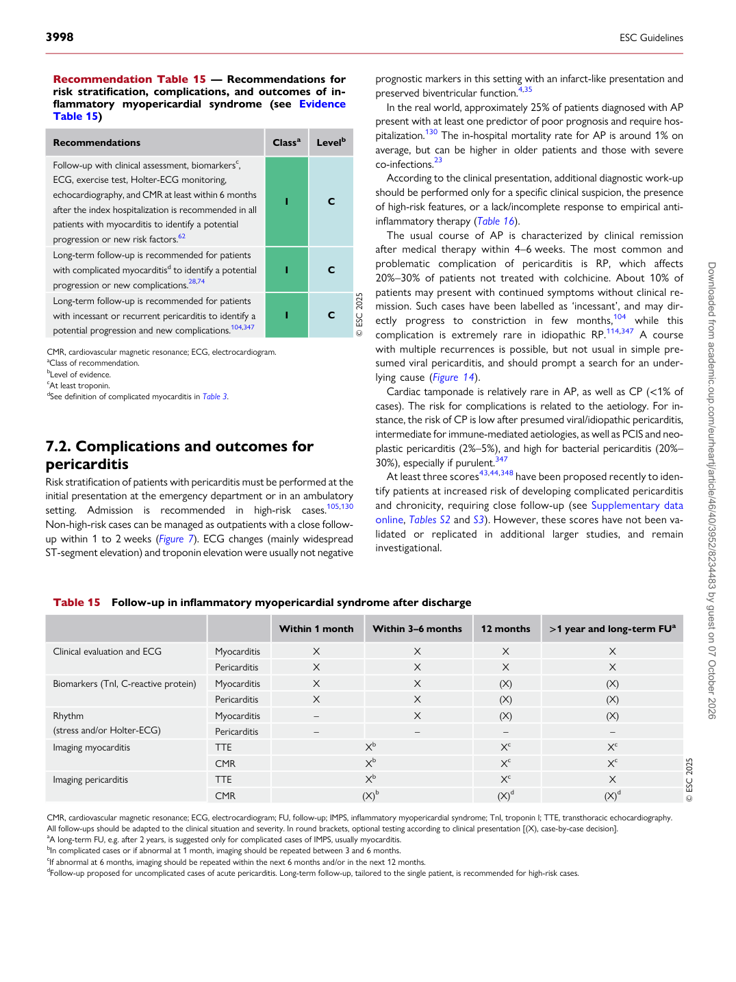
Recommendation Table 15 — Recommendations for risk stratification, complications, and outcomes of inflammatory myopericardial syndrome (see Evidence Table 15)

Recommendations Classa Levelb Follow-up with clinical assessment, biomarkersc, ECG, exercise test, Holter-ECG monitoring, echocardiography, and CMR at least within 6 months after the index hospitalization is recommended in all patients with myocarditis to identify a potential progression or new risk factors.62 I C Long-term follow-up is recommended for patients with complicated myocarditisd to identify a potential progression or new complications.28,74 I C Long-term follow-up is recommended for patients with incessant or recurrent pericarditis to identify a potential progression and new complications.104,347 I C © ESC 2025

CMR, cardiovascular magnetic resonance; ECG, electrocardiogram. aClass of recommendation. bLevel of evidence. cAt least troponin. dSee definition of complicated myocarditis in Table 3.

Table 15 Follow-up in inflammatory myopericardial syndrome after discharge

Within 1 month Within 3–6 months 12 months >1 year and long-term FUa

Clinical evaluation and ECG Myocarditis X X X X

Pericarditis X X X X

Biomarkers (TnI, C-reactive protein) Myocarditis X X (X) (X)

Pericarditis X X (X) (X)

Rhythm (stress and/or Holter-ECG) Myocarditis – X (X) (X)

Pericarditis – – – –

Imaging myocarditis TTE Xb Xc Xc

CMR Xb Xc Xc

Imaging pericarditis TTE Xb Xc X

CMR (X)b (X)d (X)d © ESC 2025

CMR, cardiovascular magnetic resonance; ECG, electrocardiogram; FU, follow-up; IMPS, inflammatory myopericardial syndrome; TnI, troponin I; TTE, transthoracic echocardiography. All follow-ups should be adapted to the clinical situation and severity. In round brackets, optional testing according to clinical presentation [(X), case-by-case decision]. aA long-term FU, e.g. after 2 years, is suggested only for complicated cases of IMPS, usually myocarditis. bIn complicated cases or if abnormal at 1 month, imaging should be repeated between 3 and 6 months. cIf abnormal at 6 months, imaging should be repeated within the next 6 months and/or in the next 12 months. dFollow-up proposed for uncomplicated cases of acute pericarditis. Long-term follow-up, tailored to the single patient, is recommended for high-risk cases. Downloaded from academic.oup.com/eurheartj/article/46/40/3952/8234483 by guest on 07 October 2026