p.36 抽出テキスト
For pericarditis, specific indications for cardiac catheterization include the differential diagnosis between restrictive CMP (RCM) or CP,252 when this differentiation cannot be done by multimodality imaging (see Supplementary data online, Section 2.6).

5.12 Electro-anatomical mapping

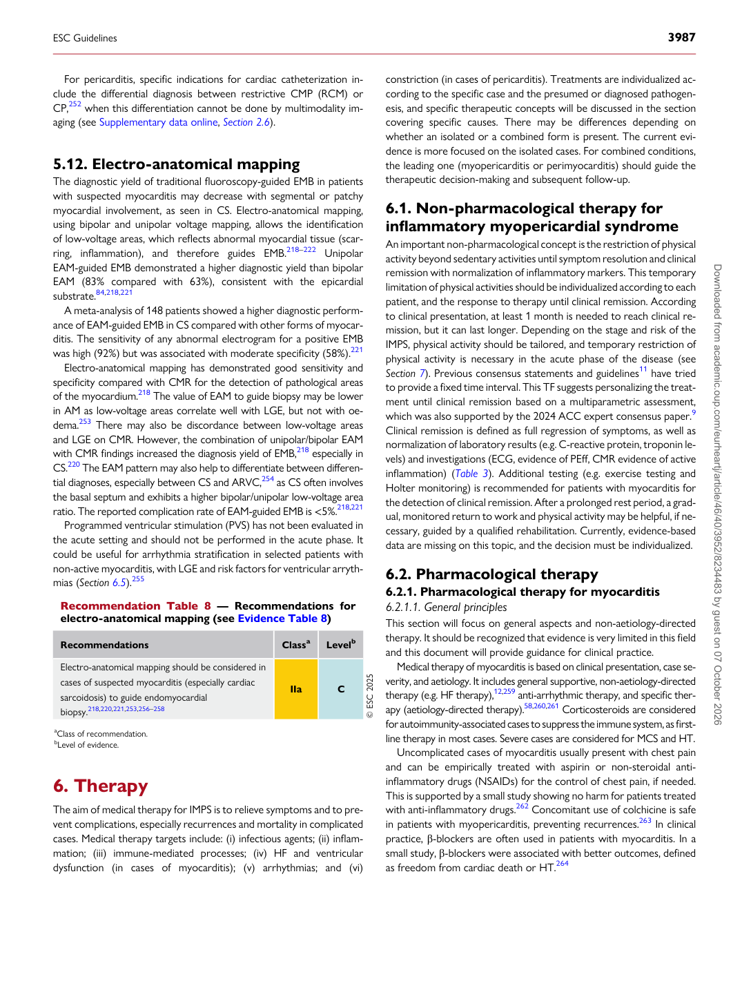
The diagnostic yield of traditional fluoroscopy-guided EMB in patients with suspected myocarditis may decrease with segmental or patchy myocardial involvement, as seen in CS. Electro-anatomical mapping, using bipolar and unipolar voltage mapping, allows the identification of low-voltage areas, which reflects abnormal myocardial tissue (scarring, inflammation), and therefore guides EMB.218–222 Unipolar EAM-guided EMB demonstrated a higher diagnostic yield than bipolar EAM (83% compared with 63%), consistent with the epicardial substrate.84,218,221 A meta-analysis of 148 patients showed a higher diagnostic performance of EAM-guided EMB in CS compared with other forms of myocarditis. The sensitivity of any abnormal electrogram for a positive EMB was high (92%) but was associated with moderate specificity (58%).221 Electro-anatomical mapping has demonstrated good sensitivity and specificity compared with CMR for the detection of pathological areas of the myocardium.218 The value of EAM to guide biopsy may be lower in AM as low-voltage areas correlate well with LGE, but not with oedema.253 There may also be discordance between low-voltage areas and LGE on CMR. However, the combination of unipolar/bipolar EAM with CMR findings increased the diagnosis yield of EMB,218 especially in CS.220 The EAM pattern may also help to differentiate between differential diagnoses, especially between CS and ARVC,254 as CS often involves the basal septum and exhibits a higher bipolar/unipolar low-voltage area ratio. The reported complication rate of EAM-guided EMB is <5%.218,221 Programmed ventricular stimulation (PVS) has not been evaluated in the acute setting and should not be performed in the acute phase. It could be useful for arrhythmia stratification in selected patients with non-active myocarditis, with LGE and risk factors for ventricular arrythmias (Section 6.5).255

6 Therapy

The aim of medical therapy for IMPS is to relieve symptoms and to prevent complications, especially recurrences and mortality in complicated cases. Medical therapy targets include: (i) infectious agents; (ii) inflammation; (iii) immune-mediated processes; (iv) HF and ventricular dysfunction (in cases of myocarditis); (v) arrhythmias; and (vi) constriction (in cases of pericarditis). Treatments are individualized according to the specific case and the presumed or diagnosed pathogenesis, and specific therapeutic concepts will be discussed in the section covering specific causes. There may be differences depending on whether an isolated or a combined form is present. The current evidence is more focused on the isolated cases. For combined conditions, the leading one (myopericarditis or perimyocarditis) should guide the therapeutic decision-making and subsequent follow-up.

6.1 Non-pharmacological therapy for

inflammatory myopericardial syndrome An important non-pharmacological concept is the restriction of physical activity beyond sedentary activities until symptom resolution and clinical remission with normalization of inflammatory markers. This temporary limitation of physical activities should be individualized according to each patient, and the response to therapy until clinical remission. According to clinical presentation, at least 1 month is needed to reach clinical remission, but it can last longer. Depending on the stage and risk of the IMPS, physical activity should be tailored, and temporary restriction of physical activity is necessary in the acute phase of the disease (see Section 7). Previous consensus statements and guidelines11 have tried to provide a fixed time interval. This TF suggests personalizing the treatment until clinical remission based on a multiparametric assessment, which was also supported by the 2024 ACC expert consensus paper.9 Clinical remission is defined as full regression of symptoms, as well as normalization of laboratory results (e.g. C-reactive protein, troponin levels) and investigations (ECG, evidence of PEff, CMR evidence of active inflammation) (Table 3). Additional testing (e.g. exercise testing and Holter monitoring) is recommended for patients with myocarditis for the detection of clinical remission. After a prolonged rest period, a gradual, monitored return to work and physical activity may be helpful, if necessary, guided by a qualified rehabilitation. Currently, evidence-based data are missing on this topic, and the decision must be individualized.

6.2 Pharmacological therapy

6.2.1. Pharmacological therapy for myocarditis 6.2.1.1. General principles This section will focus on general aspects and non-aetiology-directed therapy. It should be recognized that evidence is very limited in this field and this document will provide guidance for clinical practice. Medical therapy of myocarditis is based on clinical presentation, case severity, and aetiology. It includes general supportive, non-aetiology-directed therapy (e.g. HF therapy),12,259 anti-arrhythmic therapy, and specific therapy (aetiology-directed therapy).58,260,261 Corticosteroids are considered for autoimmunity-associated cases to suppress the immune system, as firstline therapy in most cases. Severe cases are considered for MCS and HT. Uncomplicated cases of myocarditis usually present with chest pain and can be empirically treated with aspirin or non-steroidal antiinflammatory drugs (NSAIDs) for the control of chest pain, if needed. This is supported by a small study showing no harm for patients treated with anti-inflammatory drugs.262 Concomitant use of colchicine is safe in patients with myopericarditis, preventing recurrences.263 In clinical practice, β-blockers are often used in patients with myocarditis. In a small study, β-blockers were associated with better outcomes, defined as freedom from cardiac death or HT.264

Recommendation Table 8 — Recommendations for electro-anatomical mapping (see Evidence Table 8)

Recommendations Classa Levelb

Electro-anatomical mapping should be considered in cases of suspected myocarditis (especially cardiac sarcoidosis) to guide endomyocardial biopsy.218,220,221,253,256–258 IIa C © ESC 2025 aClass of recommendation. bLevel of evidence.

ESC Guidelines                                                                                                                                                                                          3987

Downloaded from academic.oup.com/eurheartj/article/46/40/3952/8234483 by guest on 07 October 2026