p.33 抽出テキスト
5.8 Nuclear medicine

Fluorodeoxyglucose uptake reflects metabolically active inflammatory cells in AM.201 Fluorodeoxyglucose positron emission tomography can be considered as an alternative in patients when CMR is unsuitable because of an irregular heartbeat or device-related artefacts.202 Simultaneous/hybrid cardiac PET-CT/CMR imaging complement each other in the assessment of myocarditis compared with either approach alone.201,203 Fluorodeoxyglucose positron emission tomography is also a powerful tool to detect and assess the extent of extracardiac involvement in sarcoidosis (see Section 9.4). Appropriate fasting before FDG-PET is a must to obtain reliable results.204 Typical preparation protocols include a high-fat and lowcarbohydrate diet for 12–24 h followed by complete fasting other than water for the 6–12-h period prior to FDG injection. The role of nuclear imaging in pericarditis is less well established. The use of FDG-PET/CT in pericarditis is generally complementary and is most often implemented in cases where CMR cannot be used.205

5.9 Endomyocardial and pericardial biopsy

5.9.1. Endomyocardial biopsy Endomyocardial biopsy enables the identification of the underlying histological subtype of cardiac inflammation.10,34,207,208 However, noninvasive imaging methods have significantly improved, allowing the diagnosis of myocarditis by CMR. Therefore, the role of EMB in clinical practice has changed. Endomyocardial biopsy remains important for high-risk patients and in some intermediate cases when the knowledge of the histological subtype and the identification of a possible viral aetiology are important for targeted therapies (Figure 15).207,209 Endomyocardial biopsy has potential complications (e.g. cardiac perforation/tamponade, thromboembolism, valvular trauma, severe arrhythmias, and death).9,210 Rates depend on the setting and centre experience and range between 0.6% and 5%,63,211 and increase up to 26% in FM cases on mechanical circulatory support (MCS).212 Children may have higher complication rates depending on age, the experience of the centre, and the site of the biopsy (LV or RV).63,213 On this basis, the clinical use of EMB should be tailored according to the risk–benefit ratio. Histological and immunohistological investigations, as well as viral genome analysis, allow the identification of viral or immune-mediated LM, EM, GCM, and sarcoidosis.33,214,215 Moreover, EMB allows the identification of the inflammatory stages in association with the degree of myocardial necrosis and fibrosis. Endomyocardial biopsy is useful in the differential diagnosis of infiltrative/storage diseases (amyloidosis, haemochromatosis, glycogenosis), different CMPs, and genetic heart diseases33,216 if noninvasive imaging is inconclusive. This approach is consistent with other international position statements.9,210 Endomyocardial biopsy can be useful in cases of chronic CMP that is progressive or unresponsive to standard therapy. When FM, GCM or EM are suspected, early EMB is required to start immunosuppressive therapy rapidly and improve outcomes.57 In GCM, EMB has high sensitivity (80%–93%) and a positive predictive value (71%), especially if performed within 2–4 weeks from symptom onset.217

In CS, EMB may have a low sensitivity (20%–30%) due to the focal nature of inflammatory lesions, but can be improved by guiding the EMB using CMR or electro-anatomical mapping (EAM).218–222 Historically, diagnosis by EMB was based on the histological Dallas criteria in HE-stained heart tissue sections revealing myocytolysis and interstitial immune cells.223 However, since there is high interobserver variability, immunohistochemical detection of lymphocytes (CD3) and macrophages (CD68) can definitely increase sensitivity for the diagnosis of myocarditis.10,224–226 A recent meta-analysis indicates that the use of immunohistochemistry can increase the detection rate of inflammation in EMB specimens to ∼51%.227 A value of ≥14 leucocytes/mm2 with T lymphocytes of ≥7 cells/mm2 has been considered as the cut-off for the diagnosis of myocarditis.10 In acute LM, the number of T lymphocytes is generally higher (>25 cells/mm2).228 Additionally, the expression of MHC class II molecules in antigen-presenting macrophages and endothelial cells is routinely observed in EMB with myocarditis.33 Endomyocardial biopsy also enables the detection of viruses, such as enteroviruses, B19V,228 and herpesviruses, e.g. EBV and HHV-6, using quantitative realtime (RT)-PCR and in situ hybridization. It is likely that advances in molecular diagnostic techniques, such as next-generation sequencing, will help to identify potential new pathogens. Positive PCR results obtained on EMB have to be accompanied by a parallel investigation of blood samples collected at the time of the EMB to exclude acute systemic virus infection. In cases of acute cardiac and systemic virus infection, immunosuppressive therapy must be avoided. The most common site of EMB is the RV septum, but occasionally LV or biventricular EMB may be needed. The decision of the EMB site should be based on the clinical indication, findings of pre-procedural imaging, and operator expertise.229 A study of 755 patients with suspected myocarditis and non-ischaemic CMP (including infiltrative and storage disorders) indicated that biventricular EMB can increase diagnostic accuracy compared with selective LV or RV EMB.211 Sampling error is the major limitation of the diagnostic accuracy of EMB. The minimal number of required EMB samples for each technique is reported in Table 11.33 It should be emphasized that PCR in EMB tissue and in situ hybridization can be used for identification of the viral agent, because viral serology concurs with the results of PCR in a minority of patients (∼4%) with myocarditis.230 With PCR, the rate of viral sequences detected in EMB specimens is up to 67%, although the rate varies widely among studies.63,228,231 The PCR tests for the detection of infectious agents must be performed in laboratories that are accredited and certified for the identification of viruses, bacteria, and parasites. The evaluation of these PCR results must be done in the context of histological/ immunohistological findings by pathologists with expertise in cardiovascular diseases. The timely communication of biopsy results is crucial for optimal therapeutic guidance. Targeted biopsies of an inflamed heart diagnosed using histology, immunohistochemistry, and molecular virology could be correlated with modern technologies, such as genomics, epigenomics, proteomics, and metabolomics to improve the diagnosis and therapy. Recommendations for EMB can be found in Recommendation Table 1.

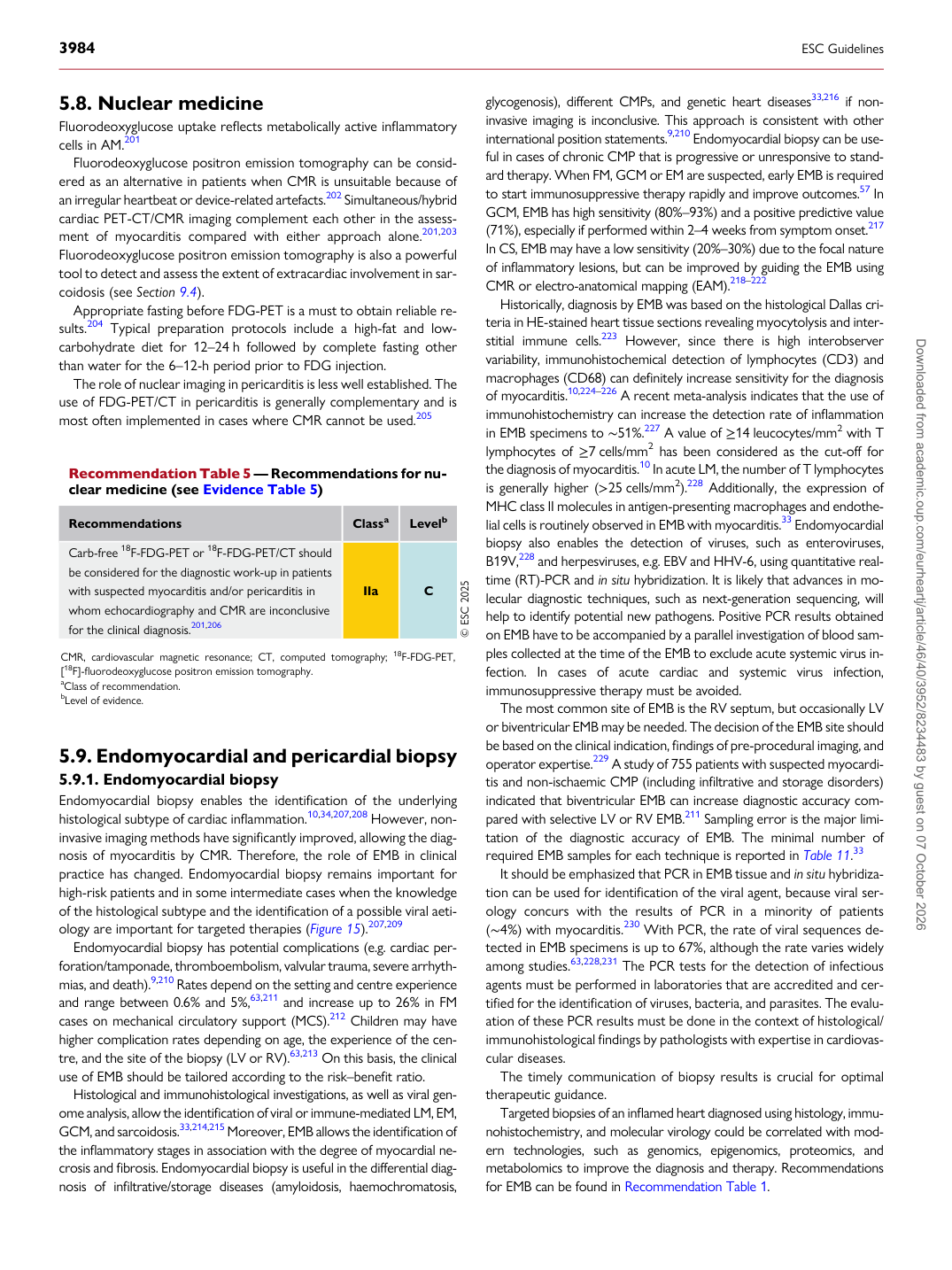
Recommendation Table 5 — Recommendations for nuclear medicine (see Evidence Table 5)

Recommendations Classa Levelb Carb-free 18F-FDG-PET or 18F-FDG-PET/CT should be considered for the diagnostic work-up in patients with suspected myocarditis and/or pericarditis in whom echocardiography and CMR are inconclusive for the clinical diagnosis.201,206 IIa C © ESC 2025 CMR, cardiovascular magnetic resonance; CT, computed tomography; 18F-FDG-PET, [18F]-fluorodeoxyglucose positron emission tomography. aClass of recommendation. bLevel of evidence. Downloaded from academic.oup.com/eurheartj/article/46/40/3952/8234483 by guest on 07 October 2026