p.32 抽出テキスト
Acute pericarditis patients may have a normal-appearing TTE (40%– 50% of cases in first episodes); however, the presence of new or worsening PEff is a diagnostic criterion for pericarditis.24,115,160 The size of PEff is assessed by the end-diastolic distance of the echo-free space between the epicardium and parietal pericardium (Table 10).115,128 It is recommended that images should include the extent and location of each effusion measurement, as this allows follow-up studies. Transthoracic echocardiography enables the recognition of CTP and the presence of constriction.115

5.6 Cardiovascular magnetic resonance

Cardiovascular magnetic resonance is the accepted gold standard for the quantification of biventricular function and detection of WMA. Cardiovascular magnetic resonance has the advantage of tissue differentiation, non-invasively detecting myocardial and pericardial inflammation, as well as fibrosis.30 The diagnostic targets in non-ischaemic myocardial inflammation include myocardial oedema, hyperaemia and capillary leak, and necrosis/fibrosis (see Supplementary data online, Figure S2).161–164 The diagnostic accuracy of CMR is higher if performed early in the time course of disease (best within the first 2 weeks). Myocardial oedema and changes due to inflammation lead to increased tissue water content that can be detected as increased signal intensities on T2-weighted imaging and/or T2 mapping; increased tissue freewater content will also lead to an increased signal in T1 mapping and extracellular volume (ECV) quantification. Myocyte necrosis and fibrosis can result in non-ischaemic (e.g. mid-wall, subepicardial, patchy) patterns of LGE, which do not typically follow a coronary artery distribution. The identification of myocarditis with chest pain presentation is less challenging than in HF and arrhythmic scenarios. The updated LLC, which include parametric mapping methods (T1/T2/ECV), should be applied for diagnosis, as the combined approach increases the diagnostic accuracy (see Figure 4).30 It is important to note that imaging evidence of myocardial inflammation does not provide the underlying histotype (see Supplementary data online, Figure S2). In the case of pericarditis, CMR detects thickening, oedema, and LGE of the pericardium.110,129,165 It is important to note that pericardial LGE may imply ongoing inflammation and neovascularization rather than simple scarring (see Supplementary data online, Figure S2). Cardiovascular magnetic resonance is helpful for an objective assessment of the degree of inflammatory involvement of the pericardium, either for first diagnoses or follow-up (for details see Figure 7 and Table 9). Given its physical properties, CMR is affected by ferromagnetic objects, such as valvular prosthesis and implants, resulting in artefacts. The same holds true for active MR-conditional devices, such as pacemakers, ICDs, and cardiac resynchronization therapy (CRT) pacemakers/defibrillators (P/Ds), but dedicated techniques are now available to achieve better diagnostic image quality in many cases.166 Cardiovascular magnetic resonance could also be used in patients with non-conditional devices with a clear clinical indication if other imaging modalities are not helpful. In all cases, programming of the implanted device before and after the scan is warranted, as well as patient monitoring. Even in intubated patients, CMR scans are feasible in order to detect inflammation.167 If breath-hold capacities are reduced or the patient is uncooperative, fast imaging techniques and/or motion-corrected imaging have to be applied.168 5.7 Computed tomography

Computed tomography is an established tool for excluding CAD depending on the risk score and can be used in the diagnostic work-up according to current ESC guidelines.65,72,195 Contrast-enhanced CT may be very useful to make an aetiological diagnosis in patients presenting with large PEff including tamponade.196 Computed tomography is important before pericardiectomy for constriction to evaluate the anatomy and extent of calcifications as a higher burden of pericardial calcification has been associated with a better post-surgical outcome.197 Furthermore, anatomical abnormalities, such as pectus excavatum, can be identified.198 Meanwhile, CT in combination with FDG-PET plays a crucial role in the detection of CS (Section 9.4).

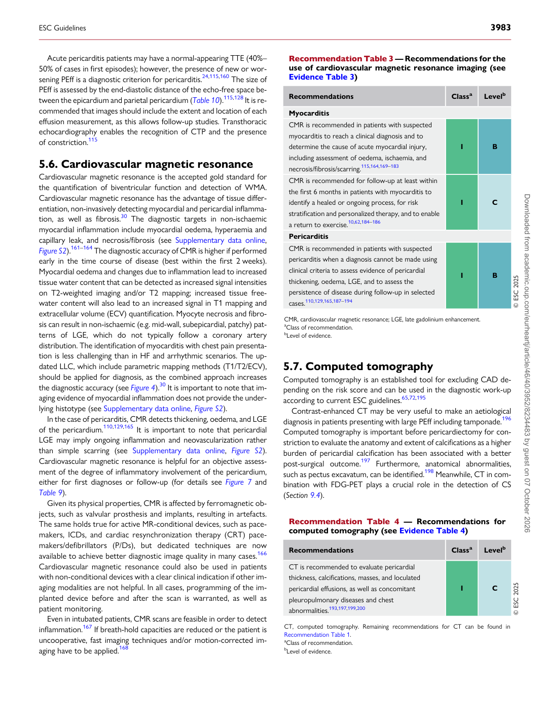
Recommendation Table 3 — Recommendations for the use of cardiovascular magnetic resonance imaging (see Evidence Table 3)

Recommendations Classa Levelb

Myocarditis CMR is recommended in patients with suspected myocarditis to reach a clinical diagnosis and to determine the cause of acute myocardial injury, including assessment of oedema, ischaemia, and necrosis/fibrosis/scarring.115,164,169–183 I B CMR is recommended for follow-up at least within the first 6 months in patients with myocarditis to identify a healed or ongoing process, for risk stratification and personalized therapy, and to enable a return to exercise.10,62,184–186 I C Pericarditis CMR is recommended in patients with suspected pericarditis when a diagnosis cannot be made using clinical criteria to assess evidence of pericardial thickening, oedema, LGE, and to assess the persistence of disease during follow-up in selected cases.110,129,165,187–194 I B © ESC 2025

CMR, cardiovascular magnetic resonance; LGE, late gadolinium enhancement. aClass of recommendation. bLevel of evidence.

Recommendation Table 4 — Recommendations for computed tomography (see Evidence Table 4)

Recommendations Classa Levelb

CT is recommended to evaluate pericardial thickness, calcifications, masses, and loculated pericardial effusions, as well as concomitant pleuropulmonary diseases and chest abnormalities.193,197,199,200 I C © ESC 2025

CT, computed tomography. Remaining recommendations for CT can be found in Recommendation Table 1. aClass of recommendation. bLevel of evidence.

ESC Guidelines                                                                                                                                                                                          3983

Downloaded from academic.oup.com/eurheartj/article/46/40/3952/8234483 by guest on 07 October 2026