p.29 抽出テキスト
5.1 Electrocardiogram

Dynamic changes in the ST segment are classic hallmarks of pericarditis and myocarditis. The most typical pattern includes widespread ST-segment elevation, reported in 20%–60% of cases (see Supplementary data online, Table S4). In pericarditis, ECG changes suggest concomitant myocardial involvement, and are indicative of inflammation of the subepicardium.35 These changes are more common in the initial phase and may be more frequent in younger patients. ST-segment depression and T-wave inversion may also be present, albeit less frequently than in patients with acute myocardial ischaemia,132 and may be related to the extent of myocardial damage.133,134 Diffuse depression of the PR segment is a classic early finding in AP.

A decrease in the amplitude of QRS complexes on ECG can be observed in cases of AP and/or AM, especially with large effusions or extensive myocardial injury. Electrical alternans of the QRS complexes has been reported, attributed to fluctuation of the heart within a large effusion (swinging heart). Lengthening of the QT interval might occur in AM. Atrial fibrillation can occur in AP, especially in predisposed patients, in whom pericardial inflammation may be a trigger for supraventricular arrhythmias.77 In contrast, VA are more common in myocarditis. AVBs and intraventricular conduction defects, particularly right bundle branch blocks, are not uncommon (see Section 4.3.3, and Supplementary data online, Table S4) and may be associated with more severe presentations.135–137 Electrocardiogram changes are highly dynamic and may evolve over a span of hours or days, with temporal progression varying significantly among patients. Typically, ST changes resolve; however, it is important to note that a normal ECG at presentation does not exclude the possibility of IMPS.

5.2 Biomarkers

5.2.1. Biomarkers in clinical routine Recommended biomarkers in IMPS include: • Markers of inflammation (e.g. C-reactive protein): C-reactive protein is often increased in patients with pericarditis (up to 80%) and AM (up to 80%), and contributes to the diagnosis.28,36,138

Negative results, however, do not exclude the diagnosis. In pericarditis, C-reactive protein is a marker of disease activity that could be used to guide the duration of anti-inflammatory therapy.36 D-dimers are currently being investigated for their diagnostic and prognostic relevance.36,139,140 • Markers of myocardial lesions (e.g. troponin): Troponin is elevated in myocardial involvement, usually indicating cardiomyocyte necrosis. Assessment of baseline troponin is crucial to diagnose the severity of myocarditis. It is typically increased in infarct-like myocarditis. • Markers of HF (BNP or NT-proBNP): Assessment of baseline level is recommended among patients with suspected myocarditis. This measurement can be repeated at discharge and during follow-up in an outpatient setting to assess the risk of clinical events. NT-proBNP can be used in patients with myocarditis as a predictive marker of HF among patients with normal or impaired LVEF.12

5.2.2 Biomarkers beyond clinical routine

Different biomarkers may help to identify a specific aetiology and to guide risk stratification. Most of them are not part of clinical routine,

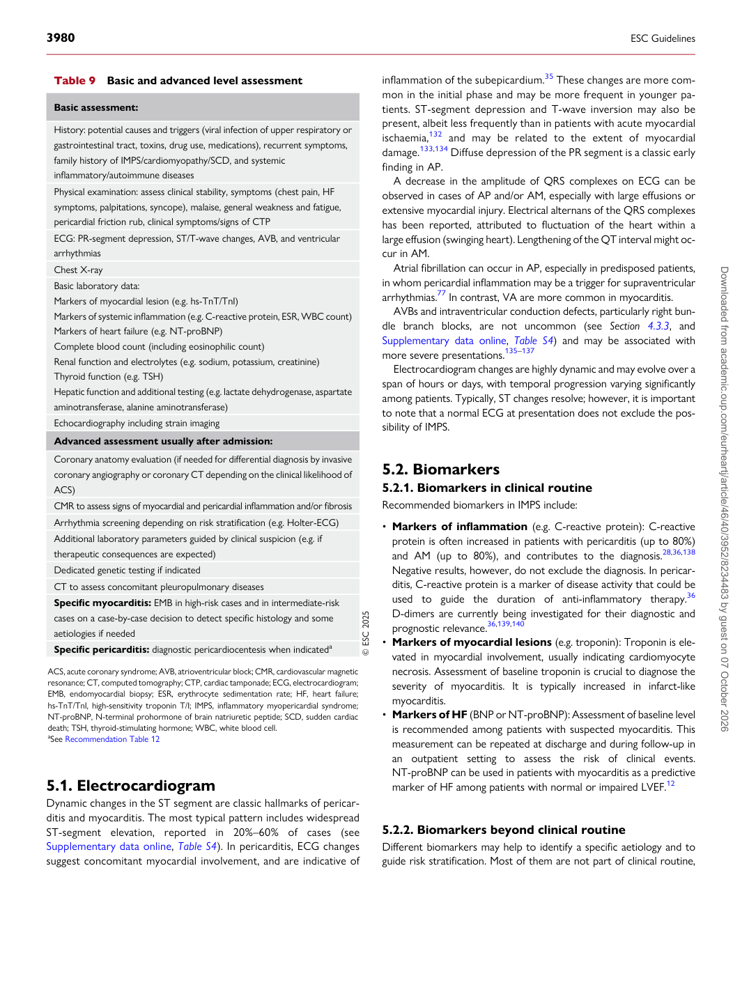
Table 9 Basic and advanced level assessment

Basic assessment:

History: potential causes and triggers (viral infection of upper respiratory or gastrointestinal tract, toxins, drug use, medications), recurrent symptoms, family history of IMPS/cardiomyopathy/SCD, and systemic inflammatory/autoimmune diseases Physical examination: assess clinical stability, symptoms (chest pain, HF symptoms, palpitations, syncope), malaise, general weakness and fatigue, pericardial friction rub, clinical symptoms/signs of CTP ECG: PR-segment depression, ST/T-wave changes, AVB, and ventricular arrhythmias Chest X-ray Basic laboratory data: Markers of myocardial lesion (e.g. hs-TnT/TnI) Markers of systemic inflammation (e.g. C-reactive protein, ESR, WBC count) Markers of heart failure (e.g. NT-proBNP) Complete blood count (including eosinophilic count) Renal function and electrolytes (e.g. sodium, potassium, creatinine) Thyroid function (e.g. TSH) Hepatic function and additional testing (e.g. lactate dehydrogenase, aspartate aminotransferase, alanine aminotransferase) Echocardiography including strain imaging Advanced assessment usually after admission: Coronary anatomy evaluation (if needed for differential diagnosis by invasive coronary angiography or coronary CT depending on the clinical likelihood of ACS) CMR to assess signs of myocardial and pericardial inflammation and/or fibrosis Arrhythmia screening depending on risk stratification (e.g. Holter-ECG) Additional laboratory parameters guided by clinical suspicion (e.g. if therapeutic consequences are expected) Dedicated genetic testing if indicated CT to assess concomitant pleuropulmonary diseases Specific myocarditis: EMB in high-risk cases and in intermediate-risk cases on a case-by-case decision to detect specific histology and some aetiologies if needed Specific pericarditis: diagnostic pericardiocentesis when indicateda © ESC 2025

ACS, acute coronary syndrome; AVB, atrioventricular block; CMR, cardiovascular magnetic resonance; CT, computed tomography; CTP, cardiac tamponade; ECG, electrocardiogram; EMB, endomyocardial biopsy; ESR, erythrocyte sedimentation rate; HF, heart failure; hs-TnT/TnI, high-sensitivity troponin T/I; IMPS, inflammatory myopericardial syndrome; NT-proBNP, N-terminal prohormone of brain natriuretic peptide; SCD, sudden cardiac death; TSH, thyroid-stimulating hormone; WBC, white blood cell. aSee Recommendation Table 12 Downloaded from academic.oup.com/eurheartj/article/46/40/3952/8234483 by guest on 07 October 2026