p.28 抽出テキスト
respiratory phase-dependent shift of the ventricular septum, and Doppler features of constriction without HF symptoms (see Supplementary data online, Section 2.4 and Figure S1).

4.5.7 Pericarditis with polyserositis

Pericarditis with polyserositis is characterized by inflammation of the pericardium and concurrent inflammation of other serous membranes lining major body cavities, such as the pleura and peritoneum.37,103,125,126

4.5.8 Inflammatory vs non-inflammatory pericarditis

In the context of pericarditis, the terms ‘inflammatory’ and ‘noninflammatory’ phenotype refer to two distinct clinical presentations based on the underlying pathophysiological mechanisms.

4.5.8.1 Inflammatory phenotype of pericarditis

The inflammatory phenotype is characterized by inflammation of the pericardium with evidence of systemic inflammation. This phenotype is often associated with fever, and/or elevated systemic inflammatory markers (e.g. C-reactive protein), PEff, and/or pleural effusion. These forms typically respond well to targeted anti-inflammatory therapies, including colchicine and anti-IL-1 agents.37,127

4.5.8.2 Non-inflammatory phenotype of pericarditis

The non-inflammatory phenotype refers to the manifestation of pericarditis without C-reactive protein elevation or low elevation of markers of systemic inflammation (e.g. high-sensitivity C-reactive protein).127 This occurs in 10%–20% of patients with pericarditis.36 The pathogenesis of pericarditis with normal C-reactive protein is unknown and these cases are often difficult to treat. C-reactive protein is a sensitive albeit unspecific biomarker of inflammation mainly driven by IL-6. Moreover, in autoimmune diseases characterized by the type I interferon (IFN) gene signature, C-reactive protein appears to be an unreliable marker of inflammation.52

5 Diagnosis and diagnostic

work-up

The diagnostic work-up in patients with suspected IMPS should be tailored based on the severity of clinical presentation, response to medical therapy, and individual risk (Table 7). It includes medical history, clinical/ physical exam, biomarkers (myocardial injury, systemic inflammation, and HF), ECG, and multimodality imaging. Endomyocardial biopsy has its place in dedicated clinical scenarios. For IMPS, routine viral serology is not recommended,1 except for HIV and hepatitis C virus (HCV), as well as for suspected bacterial infections (such as Borrelia spp., rickettsial, and T. cruzi infections). Echocardiography is the basic imaging method, followed by other techniques. Cardiovascular magnetic resonance is an advanced imaging test of increasing importance for detection of myocardial as well as pericardial injury.115,128 Most important is a patient-tailored approach, usually driven by the dominant symptoms. Flow charts (Figures 5–7 and 9–13) illustrate the proposed workflow in different clinical scenarios. If a patient presents with clinical symptoms suggestive of IMPS (Table 6), basic and advanced diagnostic assessments are recommended (see Table 9). Recommendation Table 1 — Recommendations for clinical evaluation of myocarditis and pericarditis (see Evidence Table 1)

Recommendations Classa Levelb

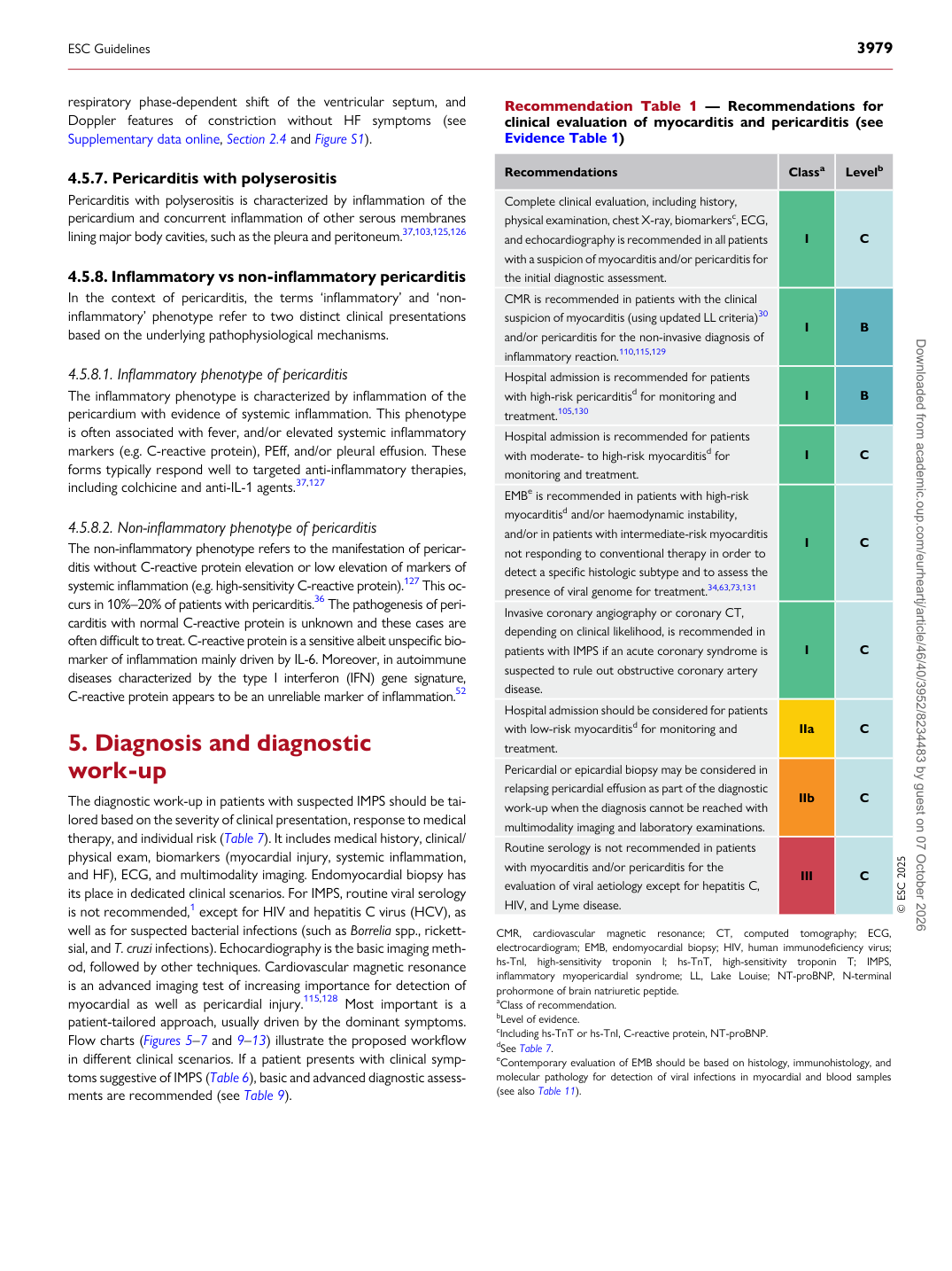
Complete clinical evaluation, including history, physical examination, chest X-ray, biomarkersc, ECG, and echocardiography is recommended in all patients with a suspicion of myocarditis and/or pericarditis for the initial diagnostic assessment. I C CMR is recommended in patients with the clinical suspicion of myocarditis (using updated LL criteria)30 and/or pericarditis for the non-invasive diagnosis of inflammatory reaction.110,115,129 I B Hospital admission is recommended for patients with high-risk pericarditisd for monitoring and treatment.105,130 I B Hospital admission is recommended for patients with moderate- to high-risk myocarditisd for monitoring and treatment. I C EMBe is recommended in patients with high-risk myocarditisd and/or haemodynamic instability, and/or in patients with intermediate-risk myocarditis not responding to conventional therapy in order to detect a specific histologic subtype and to assess the presence of viral genome for treatment.34,63,73,131 I C Invasive coronary angiography or coronary CT, depending on clinical likelihood, is recommended in patients with IMPS if an acute coronary syndrome is suspected to rule out obstructive coronary artery disease. I C Hospital admission should be considered for patients with low-risk myocarditisd for monitoring and treatment. IIa C Pericardial or epicardial biopsy may be considered in relapsing pericardial effusion as part of the diagnostic work-up when the diagnosis cannot be reached with multimodality imaging and laboratory examinations. IIb C Routine serology is not recommended in patients with myocarditis and/or pericarditis for the evaluation of viral aetiology except for hepatitis C, HIV, and Lyme disease. III C © ESC 2025

CMR, cardiovascular magnetic resonance; CT, computed tomography; ECG, electrocardiogram; EMB, endomyocardial biopsy; HIV, human immunodeficiency virus; hs-TnI, high-sensitivity troponin I; hs-TnT, high-sensitivity troponin T; IMPS, inflammatory myopericardial syndrome; LL, Lake Louise; NT-proBNP, N-terminal prohormone of brain natriuretic peptide. aClass of recommendation. bLevel of evidence. cIncluding hs-TnT or hs-TnI, C-reactive protein, NT-proBNP. dSee Table 7. eContemporary evaluation of EMB should be based on histology, immunohistology, and molecular pathology for detection of viral infections in myocardial and blood samples (see also Table 11).

ESC Guidelines                                                                                                                                                                                          3979

Downloaded from academic.oup.com/eurheartj/article/46/40/3952/8234483 by guest on 07 October 2026