p.23 抽出テキスト
Patient with signs and symptoms of acute heart failure

Clinical history and vital signs, 12-lead ECG, laboratory testinga, transthoracic echocardiography

(Class I)

Cardiogenic shock and/or respiratory failure Admission to ICU

acute Coronary syndrome Hypertensive emergency Arrhythmia Mechanical Pulmonary embolism Infections Tamponade

Further work-up Congestion/apparent HF

HF treatment

(Class I)

Red flags for myocarditisb

Suspected fulminant myocarditis

Definite myocarditis Suggestive of non cardiac causes

or cardiomyopathies Endomyocardial

biopsy (Class I)

Cardiovascular magnetic

resonancee

(Class I)

Further aetiological work-up requiredf

No further work-up

Further work-up

including geneticsg

Aetiological

work-up

Normal findings

No further work-up Risk stratificationc Before discharge

Simultaneously use CHAMPIT approach and whether congestion/apparent HF

High riskd

Non-high risk

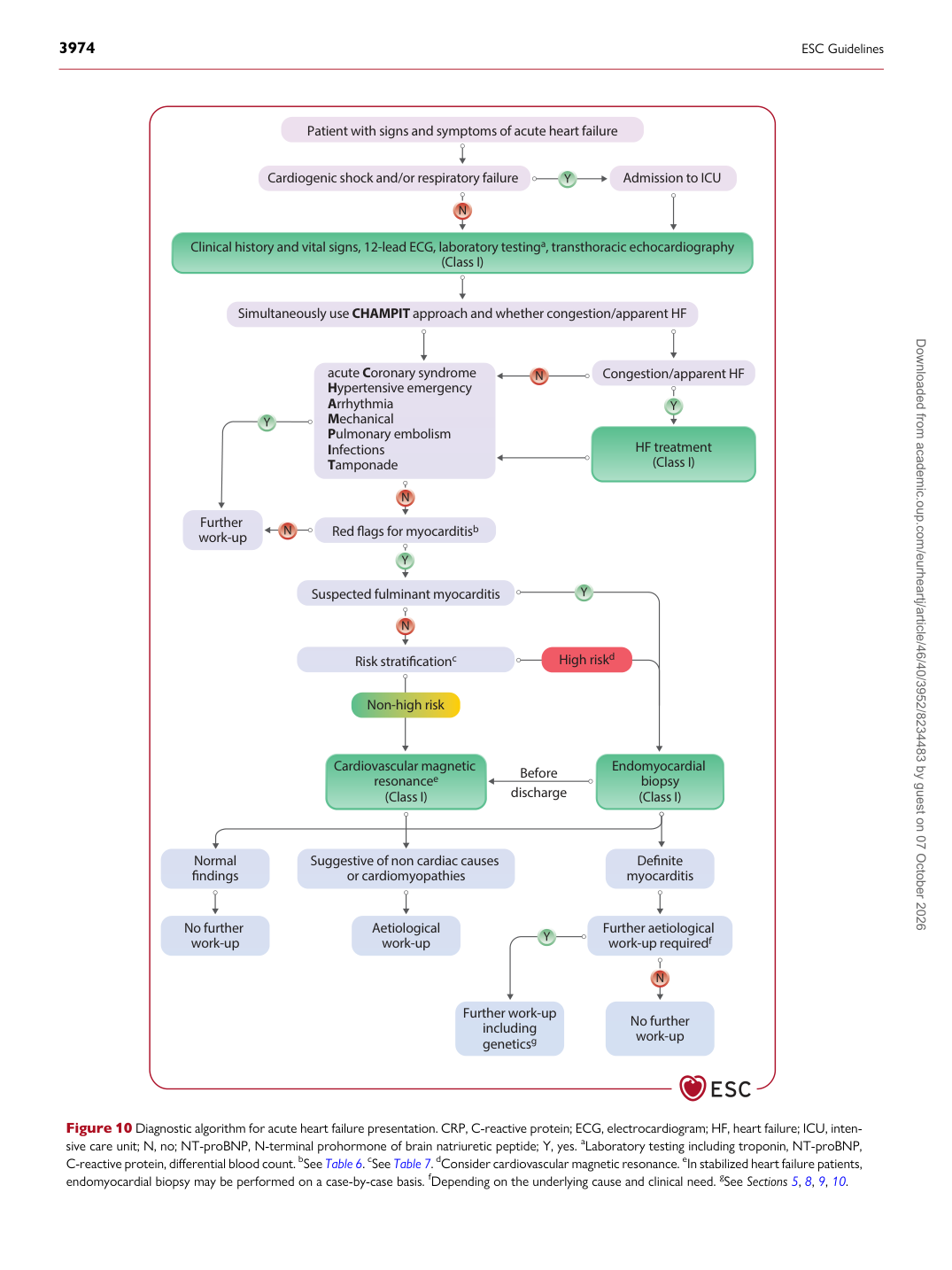
Figure 10 Diagnostic algorithm for acute heart failure presentation. CRP, C-reactive protein; ECG, electrocardiogram; HF, heart failure; ICU, intensive care unit; N, no; NT-proBNP, N-terminal prohormone of brain natriuretic peptide; Y, yes. aLaboratory testing including troponin, NT-proBNP, C-reactive protein, differential blood count. bSee Table 6. cSee Table 7. dConsider cardiovascular magnetic resonance. eIn stabilized heart failure patients, endomyocardial biopsy may be performed on a case-by-case basis. fDepending on the underlying cause and clinical need. gSee Sections 5, 8, 9, 10. Downloaded from academic.oup.com/eurheartj/article/46/40/3952/8234483 by guest on 07 October 2026