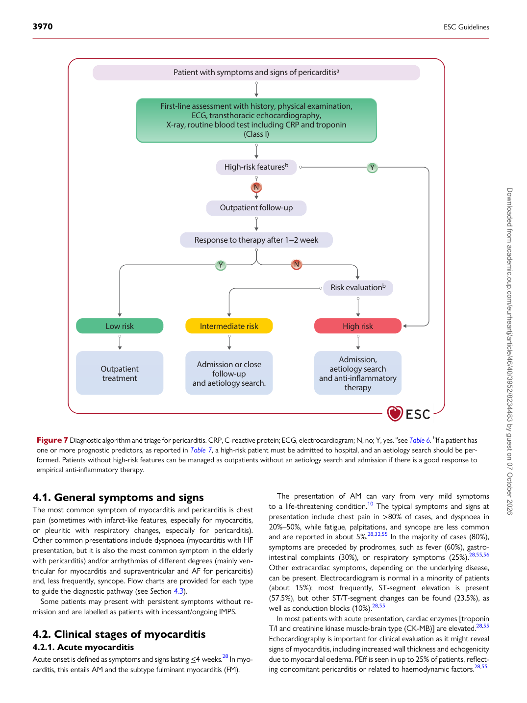
p.19 抽出テキスト
4.1 General symptoms and signs

The most common symptom of myocarditis and pericarditis is chest pain (sometimes with infarct-like features, especially for myocarditis, or pleuritic with respiratory changes, especially for pericarditis). Other common presentations include dyspnoea (myocarditis with HF presentation, but it is also the most common symptom in the elderly with pericarditis) and/or arrhythmias of different degrees (mainly ventricular for myocarditis and supraventricular and AF for pericarditis) and, less frequently, syncope. Flow charts are provided for each type to guide the diagnostic pathway (see Section 4.3). Some patients may present with persistent symptoms without remission and are labelled as patients with incessant/ongoing IMPS.

4.2 Clinical stages of myocarditis

4.2.1. Acute myocarditis Acute onset is defined as symptoms and signs lasting ≤4 weeks.28 In myocarditis, this entails AM and the subtype fulminant myocarditis (FM). The presentation of AM can vary from very mild symptoms to a life-threatening condition.10 The typical symptoms and signs at presentation include chest pain in >80% of cases, and dyspnoea in 20%–50%, while fatigue, palpitations, and syncope are less common and are reported in about 5%.28,32,55 In the majority of cases (80%), symptoms are preceded by prodromes, such as fever (60%), gastrointestinal complaints (30%), or respiratory symptoms (25%).28,55,56

Other extracardiac symptoms, depending on the underlying disease, can be present. Electrocardiogram is normal in a minority of patients (about 15%); most frequently, ST-segment elevation is present (57.5%), but other ST/T-segment changes can be found (23.5%), as well as conduction blocks (10%).28,55 In most patients with acute presentation, cardiac enzymes [troponin T/I and creatinine kinase muscle-brain type (CK-MB)] are elevated.28,55 Echocardiography is important for clinical evaluation as it might reveal signs of myocarditis, including increased wall thickness and echogenicity due to myocardial oedema. PEff is seen in up to 25% of patients, reflecting concomitant pericarditis or related to haemodynamic factors.28,55

Outpatient follow-up

Response to therapy after 1−2 week

Admission or close follow-up and aetiology search.

Patient with symptoms and signs of pericarditisa

First-line assessment with history, physical examination,

ECG, transthoracic echocardiography, X-ray, routine blood test including CRP and troponin

(Class I)

High-risk featuresb

Low risk Intermediate risk High risk

Outpatient treatment Admission, aetiology search and anti-inflammatory

therapy

Risk evaluationb

Figure 7 Diagnostic algorithm and triage for pericarditis. CRP, C-reactive protein; ECG, electrocardiogram; N, no; Y, yes. asee Table 6. bIf a patient has one or more prognostic predictors, as reported in Table 7, a high-risk patient must be admitted to hospital, and an aetiology search should be performed. Patients without high-risk features can be managed as outpatients without an aetiology search and admission if there is a good response to empirical anti-inflammatory therapy. Downloaded from academic.oup.com/eurheartj/article/46/40/3952/8234483 by guest on 07 October 2026