p.16 抽出テキスト
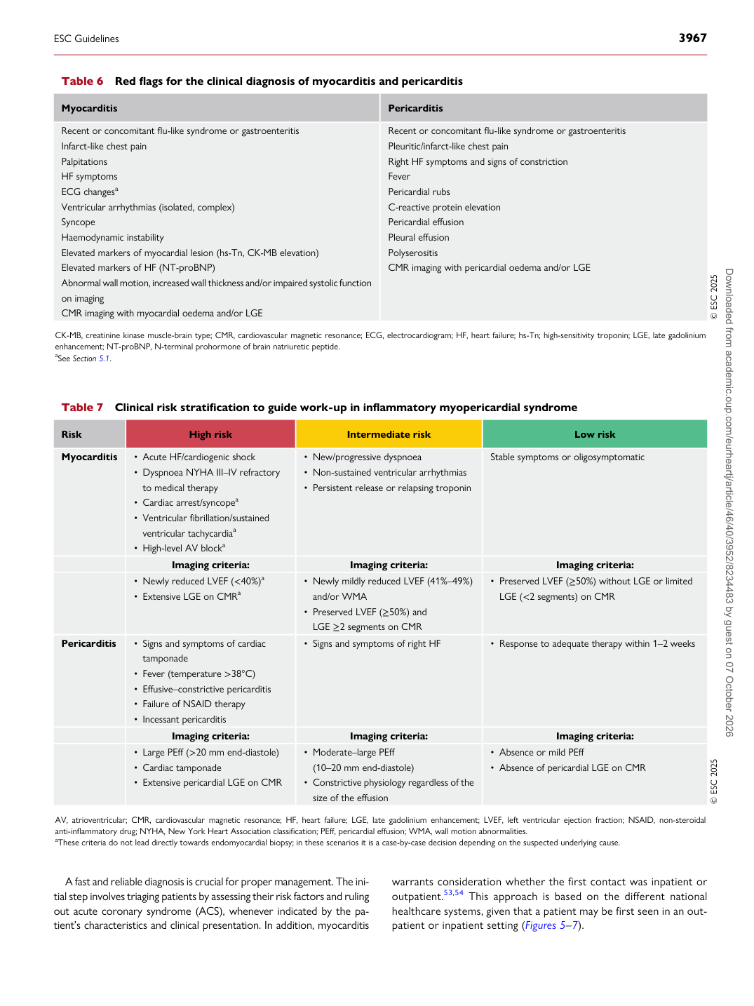
A fast and reliable diagnosis is crucial for proper management. The initial step involves triaging patients by assessing their risk factors and ruling out acute coronary syndrome (ACS), whenever indicated by the patient’s characteristics and clinical presentation. In addition, myocarditis warrants consideration whether the first contact was inpatient or outpatient.53,54 This approach is based on the different national healthcare systems, given that a patient may be first seen in an outpatient or inpatient setting (Figures 5–7).

Table 7 Clinical risk stratification to guide work-up in inflammatory myopericardial syndrome

Risk High risk Intermediate risk Low risk

Myocarditis • Acute HF/cardiogenic shock • Dyspnoea NYHA III–IV refractory

to medical therapy • Cardiac arrest/syncopea • Ventricular fibrillation/sustained

ventricular tachycardiaa • High-level AV blocka • New/progressive dyspnoea • Non-sustained ventricular arrhythmias

• Persistent release or relapsing troponin Stable symptoms or oligosymptomatic

Imaging criteria: Imaging criteria: Imaging criteria: • Newly reduced LVEF (<40%)a • Extensive LGE on CMRa • Newly mildly reduced LVEF (41%–49%)

and/or WMA • Preserved LVEF (≥50%) and LGE ≥2 segments on CMR • Preserved LVEF (≥50%) without LGE or limited

LGE (<2 segments) on CMR

Pericarditis • Signs and symptoms of cardiac tamponade

• Fever (temperature >38°C) • Effusive–constrictive pericarditis • Failure of NSAID therapy • Incessant pericarditis • Signs and symptoms of right HF • Response to adequate therapy within 1–2 weeks

Imaging criteria: Imaging criteria: Imaging criteria: • Large PEff (>20 mm end-diastole) • Cardiac tamponade • Extensive pericardial LGE on CMR • Moderate–large PEff (10–20 mm end-diastole)

• Constrictive physiology regardless of the

size of the effusion • Absence or mild PEff • Absence of pericardial LGE on CMR © ESC 2025

AV, atrioventricular; CMR, cardiovascular magnetic resonance; HF, heart failure; LGE, late gadolinium enhancement; LVEF, left ventricular ejection fraction; NSAID, non-steroidal anti-inflammatory drug; NYHA, New York Heart Association classification; PEff, pericardial effusion; WMA, wall motion abnormalities. aThese criteria do not lead directly towards endomyocardial biopsy; in these scenarios it is a case-by-case decision depending on the suspected underlying cause.

Table 6 Red flags for the clinical diagnosis of myocarditis and pericarditis

Myocarditis Pericarditis

Recent or concomitant flu-like syndrome or gastroenteritis Infarct-like chest pain Palpitations HF symptoms ECG changesa Ventricular arrhythmias (isolated, complex) Syncope Haemodynamic instability Elevated markers of myocardial lesion (hs-Tn, CK-MB elevation) Elevated markers of HF (NT-proBNP) Abnormal wall motion, increased wall thickness and/or impaired systolic function on imaging CMR imaging with myocardial oedema and/or LGE Recent or concomitant flu-like syndrome or gastroenteritis Pleuritic/infarct-like chest pain

Right HF symptoms and signs of constriction Fever Pericardial rubs C-reactive protein elevation Pericardial effusion Pleural effusion Polyserositis CMR imaging with pericardial oedema and/or LGE

© ESC 2025

CK-MB, creatinine kinase muscle-brain type; CMR, cardiovascular magnetic resonance; ECG, electrocardiogram; HF, heart failure; hs-Tn; high-sensitivity troponin; LGE, late gadolinium enhancement; NT-proBNP, N-terminal prohormone of brain natriuretic peptide. aSee Section 5.1.

ESC Guidelines                                                                                                                                                                                          3967

Downloaded from academic.oup.com/eurheartj/article/46/40/3952/8234483 by guest on 07 October 2026