p.99 抽出テキスト
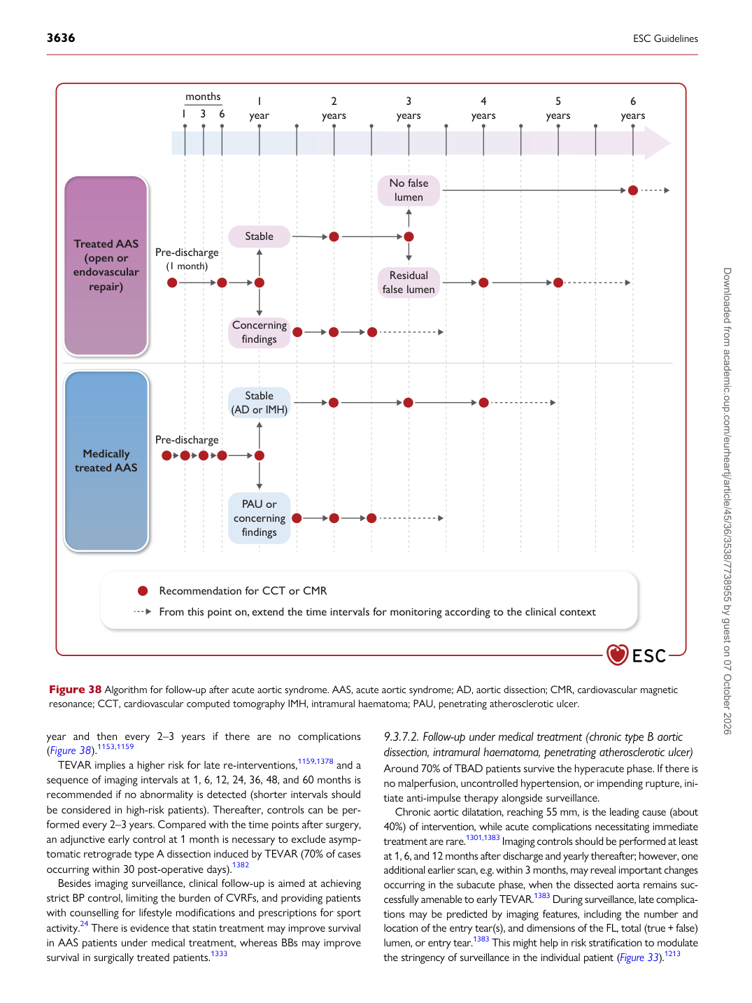
year and then every 2–3 years if there are no complications (Figure 38).1153,1159 TEVAR implies a higher risk for late re-interventions,1159,1378 and a sequence of imaging intervals at 1, 6, 12, 24, 36, 48, and 60 months is recommended if no abnormality is detected (shorter intervals should be considered in high-risk patients). Thereafter, controls can be performed every 2–3 years. Compared with the time points after surgery, an adjunctive early control at 1 month is necessary to exclude asymptomatic retrograde type A dissection induced by TEVAR (70% of cases occurring within 30 post-operative days).1382 Besides imaging surveillance, clinical follow-up is aimed at achieving strict BP control, limiting the burden of CVRFs, and providing patients with counselling for lifestyle modifications and prescriptions for sport activity.24 There is evidence that statin treatment may improve survival in AAS patients under medical treatment, whereas BBs may improve survival in surgically treated patients.1333 9.3.7.2 Follow-up under medical treatment (chronic type B aortic

dissection, intramural haematoma, penetrating atherosclerotic ulcer) Around 70% of TBAD patients survive the hyperacute phase. If there is no malperfusion, uncontrolled hypertension, or impending rupture, initiate anti-impulse therapy alongside surveillance. Chronic aortic dilatation, reaching 55 mm, is the leading cause (about 40%) of intervention, while acute complications necessitating immediate treatment are rare.1301,1383 Imaging controls should be performed at least at 1, 6, and 12 months after discharge and yearly thereafter; however, one additional earlier scan, e.g. within 3 months, may reveal important changes occurring in the subacute phase, when the dissected aorta remains successfully amenable to early TEVAR.1383 During surveillance, late complications may be predicted by imaging features, including the number and location of the entry tear(s), and dimensions of the FL, total (true + false) lumen, or entry tear.1383 This might help in risk stratification to modulate the stringency of surveillance in the individual patient (Figure 33).1213

Concerning

findings

Stable

1 6 months

3 1 year 2 years 3 years 4 years 5 years 6 years

Pre-discharge

(1 month)

Pre-discharge

Recommendation for CCT or CMR From this point on, extend the time intervals for monitoring according to the clinical context

Residual false lumen

No false lumen

Stable (AD or IMH)

PAU or concerning

findings

Treated AAS (open or endovascular

repair)

Medically treated AAS

Figure 38 Algorithm for follow-up after acute aortic syndrome. AAS, acute aortic syndrome; AD, aortic dissection; CMR, cardiovascular magnetic resonance; CCT, cardiovascular computed tomography IMH, intramural haematoma; PAU, penetrating atherosclerotic ulcer. Downloaded from academic.oup.com/eurheartj/article/45/36/3538/7738955 by guest on 07 October 2026