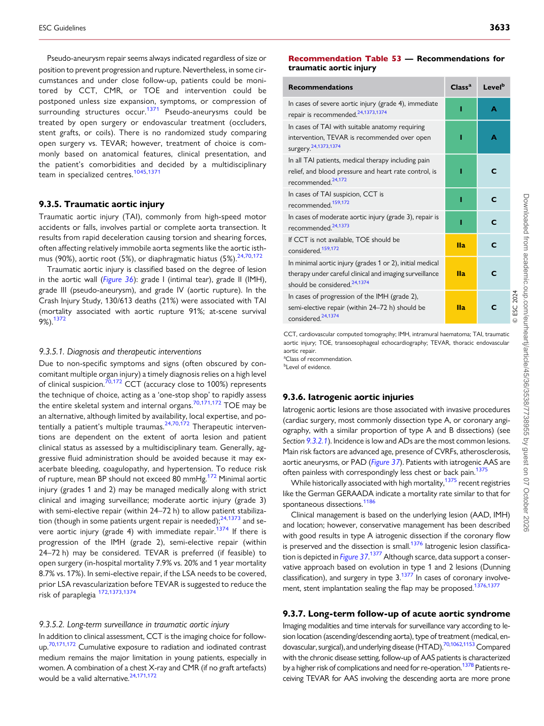
p.96 抽出テキスト
Pseudo-aneurysm repair seems always indicated regardless of size or position to prevent progression and rupture. Nevertheless, in some circumstances and under close follow-up, patients could be monitored by CCT, CMR, or TOE and intervention could be postponed unless size expansion, symptoms, or compression of surrounding structures occur.1371 Pseudo-aneurysms could be treated by open surgery or endovascular treatment (occluders, stent grafts, or coils). There is no randomized study comparing open surgery vs. TEVAR; however, treatment of choice is commonly based on anatomical features, clinical presentation, and the patient’s comorbidities and decided by a multidisciplinary team in specialized centres.1045,1371

9.3.5 Traumatic aortic injury

Traumatic aortic injury (TAI), commonly from high-speed motor accidents or falls, involves partial or complete aorta transection. It results from rapid deceleration causing torsion and shearing forces, often affecting relatively immobile aorta segments like the aortic isthmus (90%), aortic root (5%), or diaphragmatic hiatus (5%).24,70,172 Traumatic aortic injury is classified based on the degree of lesion in the aortic wall (Figure 36): grade I (intimal tear), grade II (IMH), grade III (pseudo-aneurysm), and grade IV (aortic rupture). In the Crash Injury Study, 130/613 deaths (21%) were associated with TAI (mortality associated with aortic rupture 91%; at-scene survival 9%).1372

9.3.5.1 Diagnosis and therapeutic interventions

Due to non-specific symptoms and signs (often obscured by concomitant multiple organ injury) a timely diagnosis relies on a high level of clinical suspicion.70,172 CCT (accuracy close to 100%) represents the technique of choice, acting as a ‘one-stop shop’ to rapidly assess the entire skeletal system and internal organs.70,171,172 TOE may be an alternative, although limited by availability, local expertise, and potentially a patient’s multiple traumas.24,70,172 Therapeutic interventions are dependent on the extent of aorta lesion and patient clinical status as assessed by a multidisciplinary team. Generally, aggressive fluid administration should be avoided because it may exacerbate bleeding, coagulopathy, and hypertension. To reduce risk of rupture, mean BP should not exceed 80 mmHg.172 Minimal aortic injury (grades 1 and 2) may be managed medically along with strict clinical and imaging surveillance; moderate aortic injury (grade 3) with semi-elective repair (within 24–72 h) to allow patient stabilization (though in some patients urgent repair is needed);24,1373 and severe aortic injury (grade 4) with immediate repair.1374 If there is progression of the IMH (grade 2), semi-elective repair (within 24–72 h) may be considered. TEVAR is preferred (if feasible) to open surgery (in-hospital mortality 7.9% vs. 20% and 1 year mortality 8.7% vs. 17%). In semi-elective repair, if the LSA needs to be covered, prior LSA revascularization before TEVAR is suggested to reduce the risk of paraplegia 172,1373,1374

9.3.5.2 Long-term surveillance in traumatic aortic injury

In addition to clinical assessment, CCT is the imaging choice for followup.70,171,172 Cumulative exposure to radiation and iodinated contrast medium remains the major limitation in young patients, especially in women. A combination of a chest X-ray and CMR (if no graft artefacts) would be a valid alternative.24,171,172 9.3.6 Iatrogenic aortic injuries

Iatrogenic aortic lesions are those associated with invasive procedures (cardiac surgery, most commonly dissection type A, or coronary angiography, with a similar proportion of type A and B dissections) (see Section 9.3.2.1). Incidence is low and ADs are the most common lesions. Main risk factors are advanced age, presence of CVRFs, atherosclerosis, aortic aneurysms, or PAD (Figure 37). Patients with iatrogenic AAS are often painless with correspondingly less chest or back pain.1375 While historically associated with high mortality,1375 recent registries like the German GERAADA indicate a mortality rate similar to that for spontaneous dissections.1186 Clinical management is based on the underlying lesion (AAD, IMH) and location; however, conservative management has been described with good results in type A iatrogenic dissection if the coronary flow is preserved and the dissection is small.1376 Iatrogenic lesion classification is depicted in Figure 37.1377 Although scarce, data support a conservative approach based on evolution in type 1 and 2 lesions (Dunning classification), and surgery in type 3.1377 In cases of coronary involvement, stent implantation sealing the flap may be proposed.1376,1377

9.3.7 Long-term follow-up of acute aortic syndrome

Imaging modalities and time intervals for surveillance vary according to lesion location (ascending/descending aorta), type of treatment (medical, endovascular, surgical), and underlying disease (HTAD).70,1062,1153 Compared with the chronic disease setting, follow-up of AAS patients is characterized by a higher risk of complications and need for re-operation.1378 Patients receiving TEVAR for AAS involving the descending aorta are more prone

Recommendation Table 53 — Recommendations for traumatic aortic injury

Recommendations Classa Levelb

In cases of severe aortic injury (grade 4), immediate repair is recommended.24,1373,1374 I A In cases of TAI with suitable anatomy requiring intervention, TEVAR is recommended over open surgery.24,1373,1374 I A In all TAI patients, medical therapy including pain relief, and blood pressure and heart rate control, is recommended.24,172 I C In cases of TAI suspicion, CCT is recommended.159,172 I C In cases of moderate aortic injury (grade 3), repair is recommended.24,1373 I C If CCT is not available, TOE should be considered.159,172 IIa C In minimal aortic injury (grades 1 or 2), initial medical therapy under careful clinical and imaging surveillance should be considered.24,1374 IIa C In cases of progression of the IMH (grade 2), semi-elective repair (within 24–72 h) should be considered.24,1374 IIa C © ESC 2024

CCT, cardiovascular computed tomography; IMH, intramural haematoma; TAI, traumatic aortic injury; TOE, transoesophageal echocardiography; TEVAR, thoracic endovascular aortic repair. aClass of recommendation. bLevel of evidence.

ESC Guidelines                                                                                                                                                                                          3633

Downloaded from academic.oup.com/eurheartj/article/45/36/3538/7738955 by guest on 07 October 2026