p.93 抽出テキスト
dissections, strict control of the pregnant patient and foetus with conservative medical management is recommended.1224,1335 Although limited to selected cases, successful TEVAR has been described in complicated TBAD.1227 More information is detailed in the 2018 ESC Guidelines for the management of cardiovascular diseases during pregnancy.1337

9.3.2 Intramural haematoma

Intramural haematoma, constituting 5%–25% of AAS cases, involves vasa vasorum haemorrhage within the aortic media, with or without intimal disruption (ID).70,172,1338 Most cases (60%–70%) involve the DTA (ascending aorta ∼30%, aortic arch ∼10%).70,172,1192 Although it usually occurs at an older age than AAD, risk factors and symptoms are similar;70,172,1192,1338 however, aortic regurgitation, malperfusion syndrome, and pulse deficits are less frequent in type A IMH than in TAAD.70,172

9.3.2.1 Diagnostic work-up

Diagnostic IMH work-up should be similar to that proposed for AAS (Figure 30), but with different morphological features in the imaging techniques. CCT and CMR (followed by TOE) are the leading techniques for diagnosis.70,159,171–173 Unenhanced followed by contrast-enhanced CCT represents the most used tool in the acute setting (hyperintense signal of aortic wall before contrast administration).70,171,172 The IMH diagnostic hallmark consists of crescentic or circular aortic wall thickening in the absence of an intimal flap or aortic wall enhancement following contrast administration.70,171,172 CMR is an excellent imaging technique to detect small IMHs and for the differentiation of IMH (hyperenhanced images in T1-weighted images) from atherosclerotic thickening of the aorta, thrombus, or thrombosed dissection.172 TTE yields low sensitivity (<40% for IMH cut-off limit of 5 mm).171

9.3.2.2 Clinical outcomes

Intramural haematoma may evolve into AAD (12% of patients), saccular (8%) or fusiform aneurysm (22%), and/or ID (54%).1192,1339–1342 Partial or total regression is reported in 34% of patients.70,1192,1343 Outcomes are comparable to those in AAD. In-hospital mortality for type A IMH is 26.6% (surgical 24.1% and medical 40.0%). In this regard, higher mortality for IMH involving the aortic valvular complex has been observed.1175 In-hospital mortality for type B IMH is 4.4% but worsens once surgery is indicated (surgical 20.0% vs. medical 3.8%).1175,1344

9.3.2.3 Geographical variations

Reports from South Korea and Japan reveal notable disparities with Western nations in IMH incidence (28.9% vs. 5.7% of overall AAD as reported by IRAD), treatment strategies, and outcomes. In Eastern regions, the majority (80.8%) of type A IMH patients received medical treatment, resulting in significantly improved clinical outcomes (in-hospital mortality 6.6% [5.9% for medical and 9.4% for surgical]).1345 These results may be partially explained by the detection of early-stage IMH (mild, uncomplicated cases) at primary centres.1345–1347 9.3.2.4 Management

Current IMH therapeutic interventions are similar to AAD, with the first step consisting mainly of pain and BP control regardless of the anatomopathological features (Figure 31).

9.3.2.4.1 Type A intramural haematoma. As in AAD, type A IMH

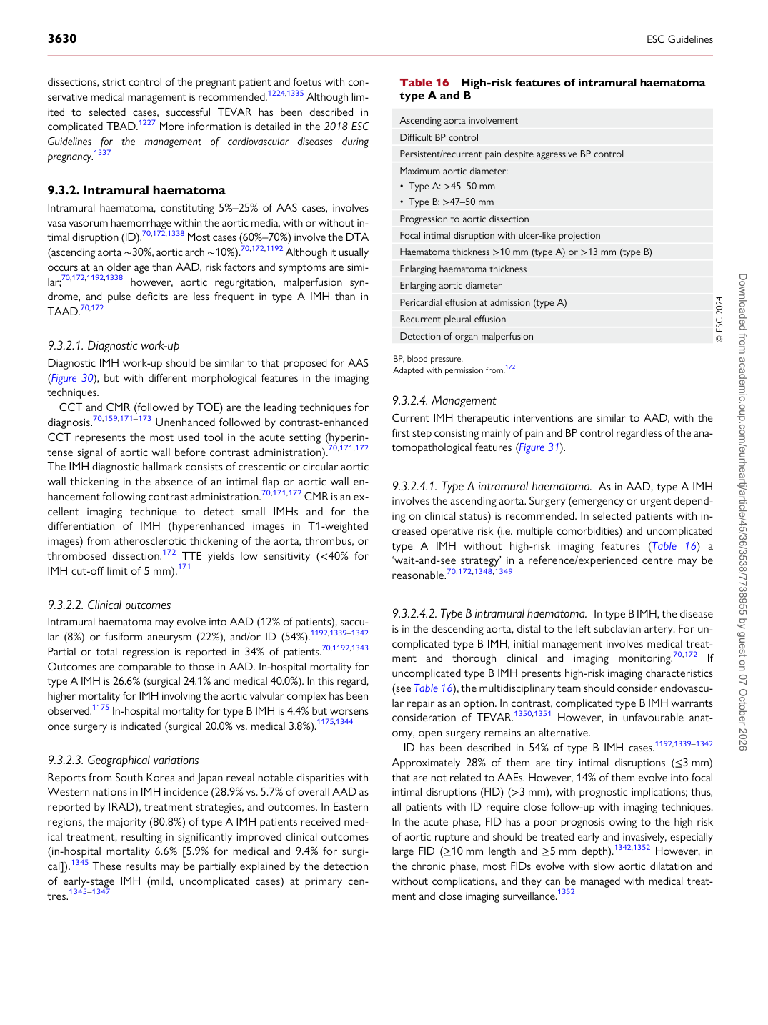
involves the ascending aorta. Surgery (emergency or urgent depending on clinical status) is recommended. In selected patients with increased operative risk (i.e. multiple comorbidities) and uncomplicated type A IMH without high-risk imaging features (Table 16) a ‘wait-and-see strategy’ in a reference/experienced centre may be reasonable.70,172,1348,1349

9.3.2.4.2 Type B intramural haematoma. In type B IMH, the disease

is in the descending aorta, distal to the left subclavian artery. For uncomplicated type B IMH, initial management involves medical treatment and thorough clinical and imaging monitoring.70,172 If uncomplicated type B IMH presents high-risk imaging characteristics (see Table 16), the multidisciplinary team should consider endovascular repair as an option. In contrast, complicated type B IMH warrants consideration of TEVAR.1350,1351 However, in unfavourable anatomy, open surgery remains an alternative.

ID has been described in 54% of type B IMH cases.1192,1339–1342 Approximately 28% of them are tiny intimal disruptions (≤3 mm) that are not related to AAEs. However, 14% of them evolve into focal intimal disruptions (FID) (>3 mm), with prognostic implications; thus, all patients with ID require close follow-up with imaging techniques. In the acute phase, FID has a poor prognosis owing to the high risk of aortic rupture and should be treated early and invasively, especially large FID (≥10 mm length and ≥5 mm depth).1342,1352 However, in the chronic phase, most FIDs evolve with slow aortic dilatation and without complications, and they can be managed with medical treatment and close imaging surveillance.1352

Table 16 High-risk features of intramural haematoma type A and B

Ascending aorta involvement Difficult BP control Persistent/recurrent pain despite aggressive BP control Maximum aortic diameter: • Type A: >45–50 mm • Type B: >47–50 mm Progression to aortic dissection Focal intimal disruption with ulcer-like projection Haematoma thickness >10 mm (type A) or >13 mm (type B) Enlarging haematoma thickness Enlarging aortic diameter Pericardial effusion at admission (type A) Recurrent pleural effusion Detection of organ malperfusion © ESC 2024

BP, blood pressure. Adapted with permission from.172 Downloaded from academic.oup.com/eurheartj/article/45/36/3538/7738955 by guest on 07 October 2026