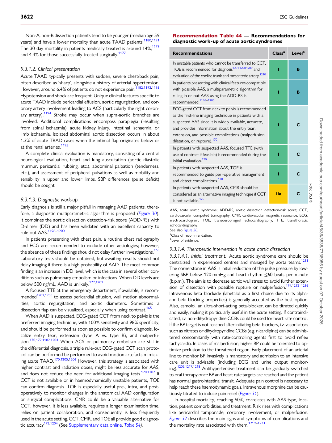
p.85 抽出テキスト
Non-A, non-B dissection patients tend to be younger (median age 59 years) and have a lower mortality than acute TAAD patients.1180,1191 The 30 day mortality in patients medically treated is around 14%,1179 and 4.4% for those successfully treated surgically.1177

9.3.1.2 Clinical presentation

Acute TAAD typically presents with sudden, severe chest/back pain, often described as ‘sharp’, alongside a history of arterial hypertension. However, around 6.4% of patients do not experience pain.1182,1192,1193 Hypotension and shock are frequent. Unique clinical features specific to acute TAAD include pericardial effusion, aortic regurgitation, and coronary artery involvement leading to ACS (particularly the right coronary artery).1194 Stroke may occur when supra-aortic branches are involved. Additional complications encompass paraplegia (resulting from spinal ischaemia), acute kidney injury, intestinal ischaemia, or limb ischaemia. Isolated abdominal aortic dissection occurs in about 1.3% of acute TBAD cases when the intimal flap originates below or at the renal arteries.1195 A complete clinical evaluation is mandatory, consisting of a central neurological evaluation, heart and lung auscultation (aortic diastolic murmur, pericardial rubbing, etc.), abdominal palpation (tenderness, etc.), and assessment of peripheral pulsations as well as mobility and sensibility in upper and lower limbs. SBP differences (pulse deficit) should be sought.

9.3.1.3 Diagnostic work-up

Early diagnosis is still a major pitfall in managing AAD patients, therefore, a diagnostic multiparametric algorithm is proposed (Figure 30). It combines the aortic dissection detection-risk score (ADD-RS) with D-dimer (DD) and has been validated with an excellent capacity to rule out AAS.1196–1200 In patients presenting with chest pain, a routine chest radiography and ECG are recommended to exclude other aetiologies; however, the absence of these findings should not delay further investigations.163 Laboratory tests should be obtained, but awaiting results should not delay imaging if there is a high probability of AAD. The most common finding is an increase in DD level, which is the case in several other conditions such as pulmonary embolism or infections. When DD levels are below 500 ng/mL, AAD is unlikely.172,1201 A focused TTE at the emergency department, if available, is recommended1202,1203 to assess pericardial effusion, wall motion abnormalities, aortic regurgitation, and aortic diameters. Sometimes a dissection flap can be visualized, especially when using contrast.165 When AAD is suspected, ECG-gated CCT from neck to pelvis is the preferred imaging technique, with 100% sensitivity and 98% specificity, and should be performed as soon as possible to confirm diagnosis, localize entry tear, extension (type A vs. type B), and malperfusion.170,172,1182,1204 When ACS or pulmonary embolism are still in the differential diagnosis, a triple rule-out ECG-gated CCT scan protocol can be performed be performed to avoid motion artefacts mimicking acute TAAD.170,1205,1206 However, this strategy is associated with higher contrast and radiation doses, might be less accurate for AAS, and does not reduce the need for additional imaging tests.170,1207 If CCT is not available or in haemodynamically unstable patients, TOE can confirm diagnosis. TOE is especially useful pre-, intra, and postoperatively to monitor changes in the anatomical AAD configuration or surgical complications. CMR could be a valuable alternative for CCT, however, it is less available, requires a longer examination time, relies on patient collaboration, and consequently, is less frequently used in the acute setting. CCT, CMR, and TOE all provide good diagnostic accuracy172,1204 (See Supplementary data online, Table S4). 9.3.1.4 Therapeutic intervention in acute aortic dissection

9.3.1.4.1. Initial treatment. Acute aortic syndrome care should be centralized in experienced centres and managed by aorta teams.1211 The cornerstone in AAS is initial reduction of the pulse pressure by lowering SBP below 120 mmHg and heart rhythm ≤60 beats per minute (b.p.m.). The aim is to decrease aortic wall stress to avoid further extension of dissection with possible rupture or malperfusion.174,1212–1216 Intravenous beta blockade (labetalol as a first choice due to its alphaand beta-blocking properties) is generally accepted as the best option. Also, esmolol, an ultra-short-acting beta-blocker, can be titrated quickly and easily, making it particularly useful in the acute setting. If contraindicated, i.v. non-dihydropyridine CCBs could be used for heart rate control. If the BP target is not reached after initiating beta-blockers, i.v. vasodilators such as nitrates or dihydropyridine CCBs (e.g. nicardipine) can be administered concomitantly with rate-controlling agents first to avoid reflex tachycardia. In cases of malperfusion, higher BP could be tolerated to optimize perfusion to the threatened region. Early placement of an arterial line to monitor BP invasively is mandatory and admission to an intensive care unit is advisable (including ECG and urine output monitoring).1205,1217,1218 Antihypertensive treatment can be gradually switched to oral therapy once BP and heart rate targets are reached and the patient has normal gastrointestinal transit. Adequate pain control is necessary to help reach these haemodynamic goals. Intravenous morphine can be cautiously titrated to induce pain relief (Figure 31). In-hospital mortality, reaching 60%, correlates with AAS type, location, patient comorbidities, and treatment. Risk rises with complications like pericardial tamponade, coronary involvement, or malperfusion. Figure 32 describes the main signs and symptoms of complications and the mortality rate associated with them.1219–1223

Recommendation Table 44 — Recommendations for diagnostic work-up of acute aortic syndromes

Recommendations Classa Levelb

In unstable patients who cannot be transferred to CCT, TOE is recommended for diagnosis1204,1208,1209 and evaluation of the coeliac trunk and mesenteric artery.1210 I B In patients presenting with clinical features compatible with possible AAS, a multiparametric algorithm for ruling in or out AAS using the ADD-RS is recommended.1196–1200 I B ECG-gated CCT from neck to pelvis is recommended as the first-line imaging technique in patients with a suspected AAS since it is widely available, accurate, and provides information about the entry tear, extension, and possible complications (malperfusion, dilatation, or rupture).170 I C In patients with suspected AAS, focused TTE (with use of contrast if feasible) is recommended during the initial evaluation.170 I C In patients with suspected AAS, TOE is recommended to guide peri-operative management and detect complications.170 I C In patients with suspected AAS, CMR should be considered as an alternative imaging technique if CCT is not available.170 IIa C © ESC 2024

AAS, acute aortic syndrome; ADD-RS, aortic dissection detection-risk score; CCT, cardiovascular computed tomography; CMR, cardiovascular magnetic resonance; ECG, electrocardiogram; TOE, transoesophageal echocardiography; TTE, transthoracic echocardiography. See also Figure 30. aClass of recommendation. bLevel of evidence. Downloaded from academic.oup.com/eurheartj/article/45/36/3538/7738955 by guest on 07 October 2026