p.81 抽出テキスト
identification of high-risk situations (e.g. older patients, inadequate sealing, type II endoleaks, no early post-procedural shrinkage of the aneurysmal sac) for which more frequent evaluation should be planned.1161,1169,1170

Follow-up of OMT is highly important in AAA patients (Figure 7).24 Statin use after AAA repair (surgical or EVAR) is associated with decreased short- and long-term mortality.1171 In addition, surveillance for aneurysm development in other arterial locations is recommended.

9.3 Acute thoracic aortic syndromes

9.3.1. General concepts Acute aortic syndromes are life-threatening emergencies, including classic AAD, IMH, PAU, aortic pseudo-aneurysm, and traumatic aortic injuries (TAI). They involve aortic wall damage and share a dynamic, overlapping pathophysiology, clinical presentation, and diagnostic and therapeutic approaches.24,172,174,910 AAS may also be iatrogenic following open or endovascular/percutaneous procedures, or cardiac surgery.1172 To guide AAS management, several anatomical classifications have been developed, the Stanford and the DeBakey systems being the most widely used. The Stanford system classifies AAS according to whether the ascending aorta is involved (type A or DeBakey type I and type II) or not (type B or DeBakey type IIIa and type IIIb) regardless of the site of origin of the intimal tear.172,174,910,1173 This classification considers not only anatomical and treatment aspects, but also prognostic implications, since patients with DeBakey type II AAS will probably be left without structural aortic wall lesions after surgery (Figure 28). Furthermore, if time elapsed from symptom onset to diagnosis is considered, AAS can be divided into hyperacute (<24 h), acute (1–14 days), subacute (15–90 days), and chronic (>90 days) (Figure 28).1174–1176 A new classification considers the intimal tear’s entry site and dissection extension (Figure 29).136 Subscript P describes the proximal involved aorta, and subscript D indicates the distal zone. This classification guides treatment decisions for sealing the entry tear. AADs limited to the aortic arch or originating as retrograde dissections from the descending aorta that extend into the arch and stop before the ascending aorta are termed as non-A non-B AD.1177–1179 Recently, a European update of the Stanford classification—Type Entry Malperfusion (TEM) classification—has been proposed.1180 This combines information about the type of dissection, its extent, and the presence of complications (malperfusion), thus providing greater prognostic insights (Figure 29). This classification is recommended by the European Association for Cardio-Thoracic Surgery. The TEM and other classifications are described in the Supplementary data online, Section 1.6.

9.3.1.1 Epidemiology and risk factors

Classic AAD (comprising 80%–90% of AAS; incidence of 2.6–3.5 cases per 100 000 person-years)24,1181 is characterized by the presence of an intimal flap separating the true from the false lumen (FL).24,172,910

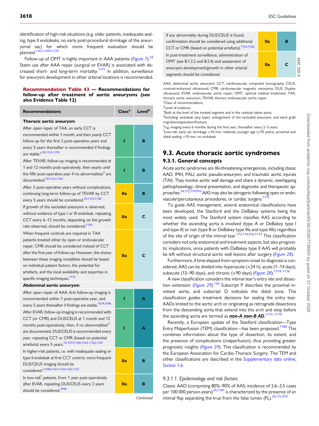
Recommendation Table 43 — Recommendations for follow-up after treatment of aortic aneurysms (see also Evidence Table 12)

Recommendations Classa Levelb

Thoracic aortic aneurysm After open repair of TAA, an early CCT is recommended within 1 month, and then yearly CCT follow-up for the first 2 post-operative years and every 5 years thereafter is recommended if findings are stable.c,70,1153,1159 I B After TEVAR, follow-up imaging is recommended at 1 and 12 months post-operatively, then yearly until the fifth post-operative year if no abnormalitiesd are documented.70,1153,1158 I B After 5 post-operative years without complications, continuing long-term follow-up of TEVAR by CCT every 5 years should be considered.70,1153,1158 IIa B If growth of the excluded aneurysm is observed, without evidence of type I or III endoleak, repeating CCT every 6–12 months, depending on the growth rate observed, should be considered.1150 IIa C When frequent controls are required in TAA patients treated either by open or endovascular repair, CMR should be considered instead of CCT after the first year of follow-up. However, the choice between these imaging modalities should be based on individual patient factors, the potential for artefacts, and the local availability and expertise in specific imaging techniques.1155 IIa C Abdominal aortic aneurysm After open repair of AAA, first follow-up imaging is recommended within 1 post-operative year, and every 5 years thereafter if findings are stable.1079,1096 I A After EVAR, follow-up imaging is recommended with CCT (or CMR) and DUS/CEUS at 1 month and 12 months post-operatively, then, if no abnormalitiesd are documented, DUS/CEUS is recommended every year, repeating CCT or CMR (based on potential artefacts) every 5 years.70,1079,1100,1163–1165,1167 I A In higher-risk patients, i.e. with inadequate sealing or type II endoleak at first CCT control, more frequent DUS/CEUS imaging should be considered.e,1096,1161,1164,1165,1167 IIa B In low-riskf patients, from 1 year post-operatively after EVAR, repeating DUS/CEUS every 2 years should be considered.1096 IIa B

Continued If any abnormality during DUS/CEUS is found,

confirmation should be considered using additional CCT or CMR (based on potential artefacts).1163,1166 IIa B In post-treatment surveillance, administration of OMT (see 8.1.2.2 and 8.2.4) and assessment of aneurysm development/growth in other arterial segments should be considered. IIa C © ESC 2024

AAA, abdominal aortic aneurysm; CCT, cardiovascular computed tomography; CEUS, contrast-enhanced ultrasound; CMR, cardiovascular magnetic resonance; DUS, Duplex ultrasound; EVAR, endovascular aortic repair; OMT, optimal medical treatment; TAA, thoracic aortic aneurysm; TEVAR, thoracic endovascular aortic repair. aClass of recommendation. bLevel of evidence. cBoth at the level of the treated segment and in the residual native aorta. dIncluding: endoleak (any type), enlargement of the excluded aneurysm, and stent graft migration/separation/fracture. ee.g. imaging every 6 months during the first year, thereafter every 2–3 years. fLow-risk: early sac shrinkage >10 mm, relatively younger age (<70 years), proximal and distal sealing >10 mm, no endoleak. Downloaded from academic.oup.com/eurheartj/article/45/36/3538/7738955 by guest on 07 October 2026