p.80 抽出テキスト
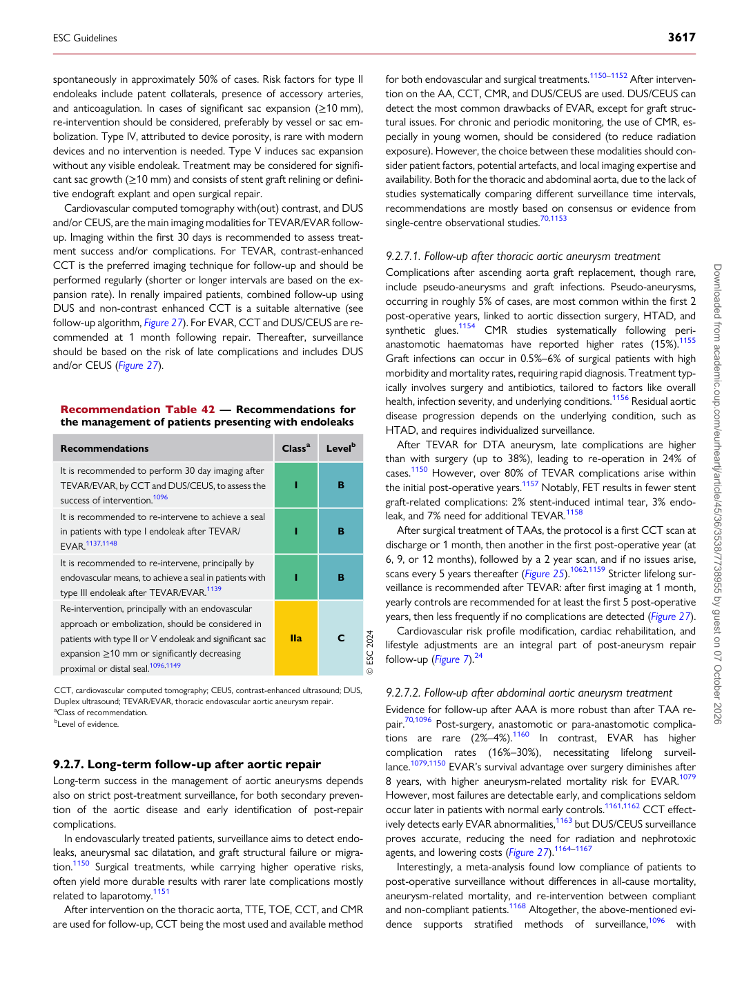
spontaneously in approximately 50% of cases. Risk factors for type II endoleaks include patent collaterals, presence of accessory arteries, and anticoagulation. In cases of significant sac expansion (≥10 mm), re-intervention should be considered, preferably by vessel or sac embolization. Type IV, attributed to device porosity, is rare with modern devices and no intervention is needed. Type V induces sac expansion without any visible endoleak. Treatment may be considered for significant sac growth (≥10 mm) and consists of stent graft relining or definitive endograft explant and open surgical repair. Cardiovascular computed tomography with(out) contrast, and DUS and/or CEUS, are the main imaging modalities for TEVAR/EVAR followup. Imaging within the first 30 days is recommended to assess treatment success and/or complications. For TEVAR, contrast-enhanced CCT is the preferred imaging technique for follow-up and should be performed regularly (shorter or longer intervals are based on the expansion rate). In renally impaired patients, combined follow-up using DUS and non-contrast enhanced CCT is a suitable alternative (see follow-up algorithm, Figure 27). For EVAR, CCT and DUS/CEUS are recommended at 1 month following repair. Thereafter, surveillance should be based on the risk of late complications and includes DUS and/or CEUS (Figure 27).

9.2.7 Long-term follow-up after aortic repair

Long-term success in the management of aortic aneurysms depends also on strict post-treatment surveillance, for both secondary prevention of the aortic disease and early identification of post-repair complications. In endovascularly treated patients, surveillance aims to detect endoleaks, aneurysmal sac dilatation, and graft structural failure or migration.1150 Surgical treatments, while carrying higher operative risks, often yield more durable results with rarer late complications mostly related to laparotomy.1151 After intervention on the thoracic aorta, TTE, TOE, CCT, and CMR are used for follow-up, CCT being the most used and available method for both endovascular and surgical treatments.1150–1152 After intervention on the AA, CCT, CMR, and DUS/CEUS are used. DUS/CEUS can detect the most common drawbacks of EVAR, except for graft structural issues. For chronic and periodic monitoring, the use of CMR, especially in young women, should be considered (to reduce radiation exposure). However, the choice between these modalities should consider patient factors, potential artefacts, and local imaging expertise and availability. Both for the thoracic and abdominal aorta, due to the lack of studies systematically comparing different surveillance time intervals, recommendations are mostly based on consensus or evidence from single-centre observational studies.70,1153

9.2.7.1 Follow-up after thoracic aortic aneurysm treatment

Complications after ascending aorta graft replacement, though rare, include pseudo-aneurysms and graft infections. Pseudo-aneurysms, occurring in roughly 5% of cases, are most common within the first 2 post-operative years, linked to aortic dissection surgery, HTAD, and synthetic glues.1154 CMR studies systematically following perianastomotic haematomas have reported higher rates (15%).1155 Graft infections can occur in 0.5%–6% of surgical patients with high morbidity and mortality rates, requiring rapid diagnosis. Treatment typically involves surgery and antibiotics, tailored to factors like overall health, infection severity, and underlying conditions.1156 Residual aortic disease progression depends on the underlying condition, such as HTAD, and requires individualized surveillance. After TEVAR for DTA aneurysm, late complications are higher than with surgery (up to 38%), leading to re-operation in 24% of cases.1150 However, over 80% of TEVAR complications arise within the initial post-operative years.1157 Notably, FET results in fewer stent graft-related complications: 2% stent-induced intimal tear, 3% endoleak, and 7% need for additional TEVAR.1158 After surgical treatment of TAAs, the protocol is a first CCT scan at discharge or 1 month, then another in the first post-operative year (at 6, 9, or 12 months), followed by a 2 year scan, and if no issues arise, scans every 5 years thereafter (Figure 25).1062,1159 Stricter lifelong surveillance is recommended after TEVAR: after first imaging at 1 month, yearly controls are recommended for at least the first 5 post-operative years, then less frequently if no complications are detected (Figure 27). Cardiovascular risk profile modification, cardiac rehabilitation, and lifestyle adjustments are an integral part of post-aneurysm repair follow-up (Figure 7).24

9.2.7.2 Follow-up after abdominal aortic aneurysm treatment

Evidence for follow-up after AAA is more robust than after TAA repair.70,1096 Post-surgery, anastomotic or para-anastomotic complications are rare (2%–4%).1160 In contrast, EVAR has higher complication rates (16%–30%), necessitating lifelong surveillance.1079,1150 EVAR’s survival advantage over surgery diminishes after 8 years, with higher aneurysm-related mortality risk for EVAR.1079 However, most failures are detectable early, and complications seldom occur later in patients with normal early controls.1161,1162 CCT effectively detects early EVAR abnormalities,1163 but DUS/CEUS surveillance proves accurate, reducing the need for radiation and nephrotoxic agents, and lowering costs (Figure 27).1164–1167 Interestingly, a meta-analysis found low compliance of patients to post-operative surveillance without differences in all-cause mortality, aneurysm-related mortality, and re-intervention between compliant and non-compliant patients.1168 Altogether, the above-mentioned evidence supports stratified methods of surveillance,1096 with

Recommendation Table 42 — Recommendations for the management of patients presenting with endoleaks

Recommendations Classa Levelb

It is recommended to perform 30 day imaging after TEVAR/EVAR, by CCT and DUS/CEUS, to assess the success of intervention.1096 I B It is recommended to re-intervene to achieve a seal in patients with type I endoleak after TEVAR/ EVAR.1137,1148 I B It is recommended to re-intervene, principally by endovascular means, to achieve a seal in patients with type III endoleak after TEVAR/EVAR.1139 I B Re-intervention, principally with an endovascular approach or embolization, should be considered in patients with type II or V endoleak and significant sac expansion ≥10 mm or significantly decreasing proximal or distal seal.1096,1149 IIa C © ESC 2024

CCT, cardiovascular computed tomography; CEUS, contrast-enhanced ultrasound; DUS, Duplex ultrasound; TEVAR/EVAR, thoracic endovascular aortic aneurysm repair. aClass of recommendation. bLevel of evidence.

ESC Guidelines                                                                                                                                                                                          3617

Downloaded from academic.oup.com/eurheartj/article/45/36/3538/7738955 by guest on 07 October 2026