p.79 抽出テキスト
Y

N Classification of endoleak types

Type Ia Type Ib Type II Type III Type IV Type V

Surveillance after TEVAR/EVAR and management of endoleaks

CCT and DUS/CEUSa at 1 month and 1 year post-op

No new endoleak Type I or type III endoleak

Consider re-intervention

(Class I)

Type II endoleak

Detected at 1 month post-op

CCT re-evaluation at 6–12 months

Annual surveillance with CCT or DUS/CEUSa for the first 5 years <10 mm or shrinking Consider open surgery, if growing aneurysm sac

CCT or DUS/CEUSa surveillance every 6 months

for 24 months

New endoleak or growing aneurysm sac

CCT, or non-contrast CCT with DUS/CEUS every 5 yearsa Growing aneurysm sac Consider embolization,

if feasible (Class IIa)

CCT

Proceed to endoleak type CCT or DUS/CEUSa

surveillance at 6–12 months

New endoleak ≥10 mm

Y

N

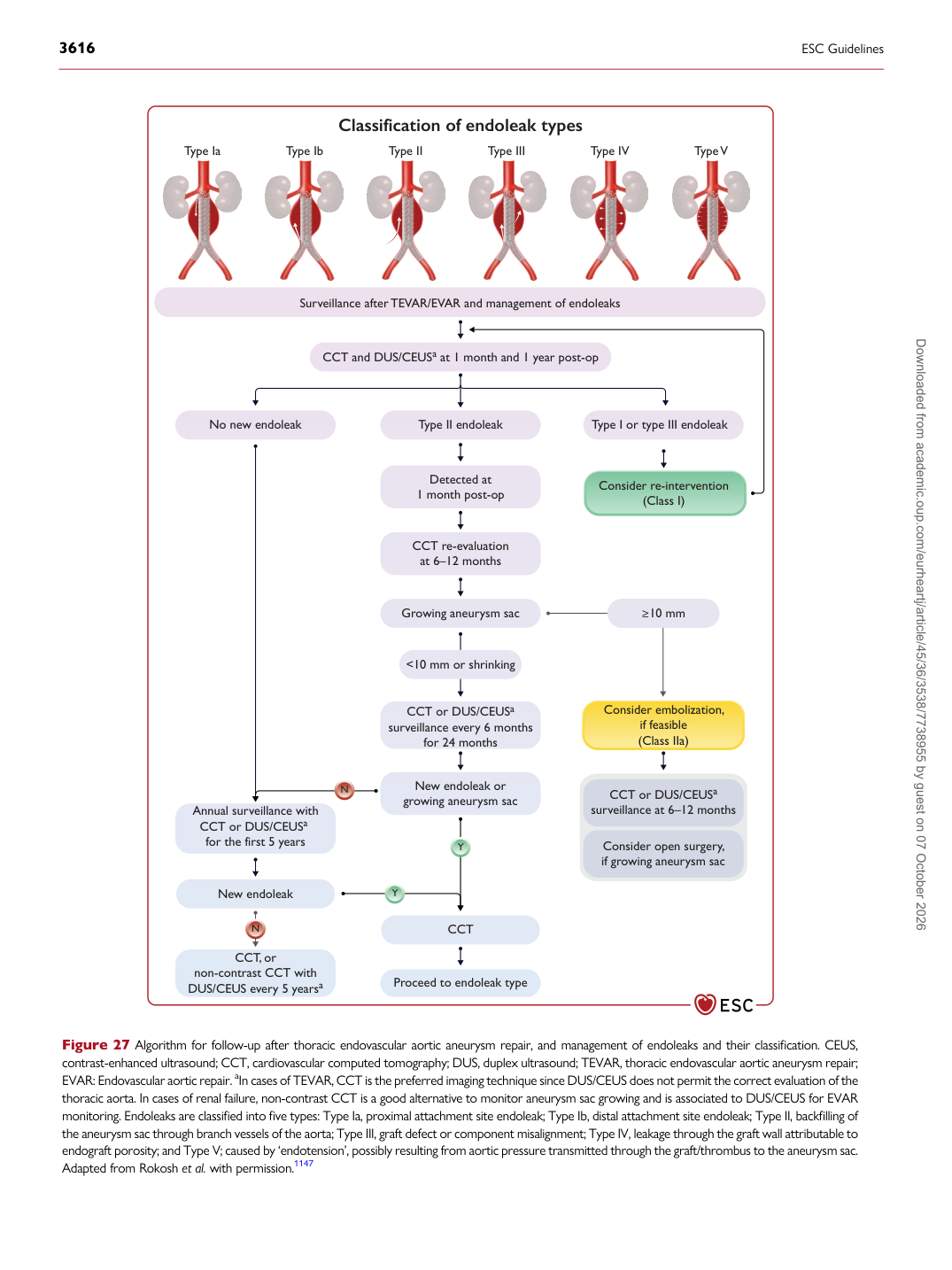
Figure 27 Algorithm for follow-up after thoracic endovascular aortic aneurysm repair, and management of endoleaks and their classification. CEUS, contrast-enhanced ultrasound; CCT, cardiovascular computed tomography; DUS, duplex ultrasound; TEVAR, thoracic endovascular aortic aneurysm repair; EVAR: Endovascular aortic repair. aIn cases of TEVAR, CCT is the preferred imaging technique since DUS/CEUS does not permit the correct evaluation of the thoracic aorta. In cases of renal failure, non-contrast CCT is a good alternative to monitor aneurysm sac growing and is associated to DUS/CEUS for EVAR monitoring. Endoleaks are classified into five types: Type Ia, proximal attachment site endoleak; Type Ib, distal attachment site endoleak; Type II, backfilling of the aneurysm sac through branch vessels of the aorta; Type III, graft defect or component misalignment; Type IV, leakage through the graft wall attributable to endograft porosity; and Type V; caused by ‘endotension’, possibly resulting from aortic pressure transmitted through the graft/thrombus to the aneurysm sac. Adapted from Rokosh et al. with permission.1147 Downloaded from academic.oup.com/eurheartj/article/45/36/3538/7738955 by guest on 07 October 2026