p.70 抽出テキスト
9.2.2.4 Surveillance

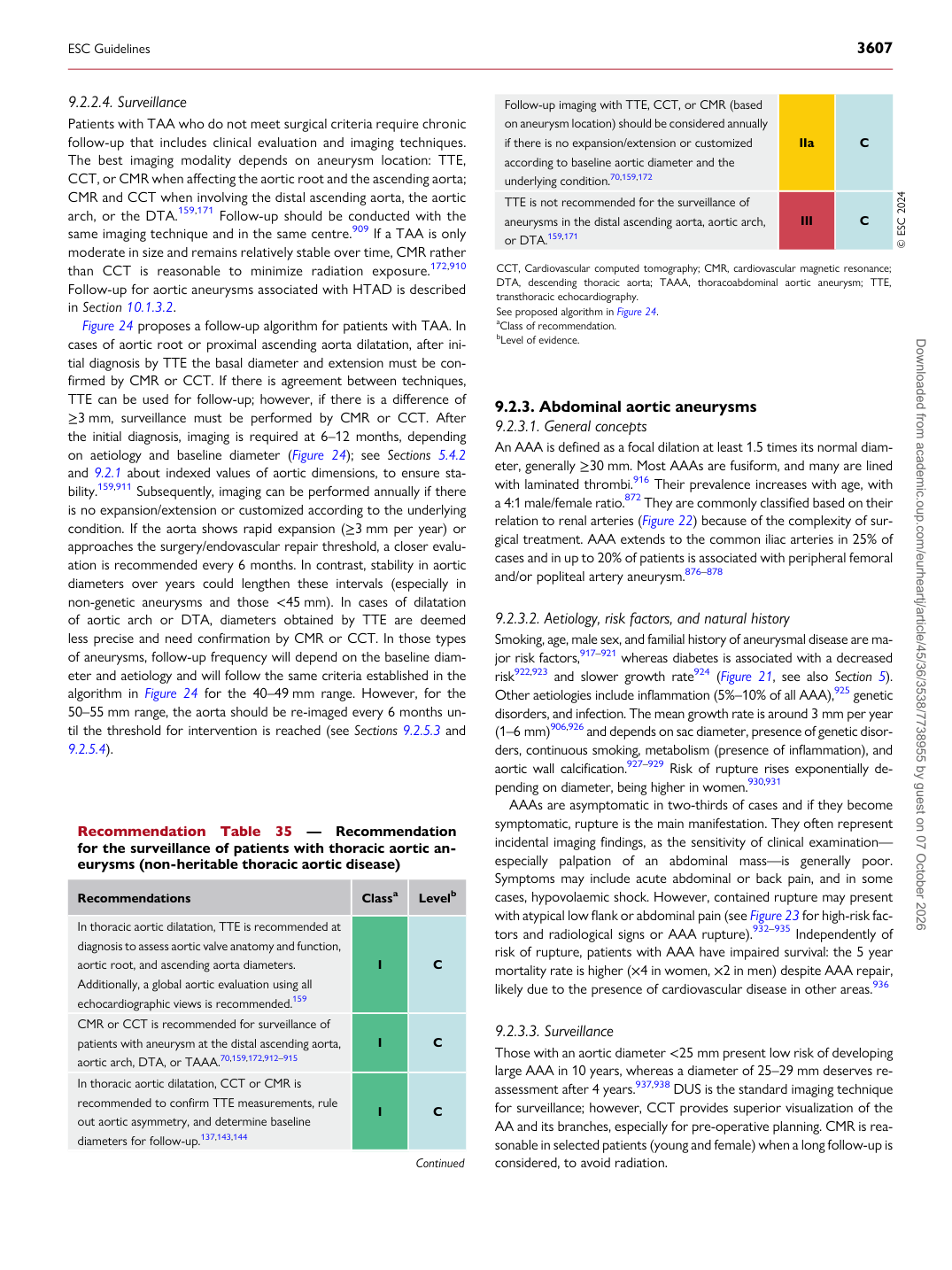
Patients with TAA who do not meet surgical criteria require chronic follow-up that includes clinical evaluation and imaging techniques. The best imaging modality depends on aneurysm location: TTE, CCT, or CMR when affecting the aortic root and the ascending aorta; CMR and CCT when involving the distal ascending aorta, the aortic arch, or the DTA.159,171 Follow-up should be conducted with the same imaging technique and in the same centre.909 If a TAA is only moderate in size and remains relatively stable over time, CMR rather than CCT is reasonable to minimize radiation exposure.172,910 Follow-up for aortic aneurysms associated with HTAD is described in Section 10.1.3.2. Figure 24 proposes a follow-up algorithm for patients with TAA. In cases of aortic root or proximal ascending aorta dilatation, after initial diagnosis by TTE the basal diameter and extension must be confirmed by CMR or CCT. If there is agreement between techniques, TTE can be used for follow-up; however, if there is a difference of ≥3 mm, surveillance must be performed by CMR or CCT. After the initial diagnosis, imaging is required at 6–12 months, depending on aetiology and baseline diameter (Figure 24); see Sections 5.4.2 and 9.2.1 about indexed values of aortic dimensions, to ensure stability.159,911 Subsequently, imaging can be performed annually if there is no expansion/extension or customized according to the underlying condition. If the aorta shows rapid expansion (≥3 mm per year) or approaches the surgery/endovascular repair threshold, a closer evaluation is recommended every 6 months. In contrast, stability in aortic diameters over years could lengthen these intervals (especially in non-genetic aneurysms and those <45 mm). In cases of dilatation of aortic arch or DTA, diameters obtained by TTE are deemed less precise and need confirmation by CMR or CCT. In those types of aneurysms, follow-up frequency will depend on the baseline diameter and aetiology and will follow the same criteria established in the algorithm in Figure 24 for the 40–49 mm range. However, for the 50–55 mm range, the aorta should be re-imaged every 6 months until the threshold for intervention is reached (see Sections 9.2.5.3 and 9.2.5.4). 9.2.3 Abdominal aortic aneurysms

9.2.3.1. General concepts An AAA is defined as a focal dilation at least 1.5 times its normal diameter, generally ≥30 mm. Most AAAs are fusiform, and many are lined with laminated thrombi.916 Their prevalence increases with age, with a 4:1 male/female ratio.872 They are commonly classified based on their relation to renal arteries (Figure 22) because of the complexity of surgical treatment. AAA extends to the common iliac arteries in 25% of cases and in up to 20% of patients is associated with peripheral femoral and/or popliteal artery aneurysm.876–878

9.2.3.2 Aetiology, risk factors, and natural history

Smoking, age, male sex, and familial history of aneurysmal disease are major risk factors,917–921 whereas diabetes is associated with a decreased risk922,923 and slower growth rate924 (Figure 21, see also Section 5). Other aetiologies include inflammation (5%–10% of all AAA),925 genetic disorders, and infection. The mean growth rate is around 3 mm per year (1–6 mm)906,926 and depends on sac diameter, presence of genetic disorders, continuous smoking, metabolism (presence of inflammation), and aortic wall calcification.927–929 Risk of rupture rises exponentially depending on diameter, being higher in women.930,931 AAAs are asymptomatic in two-thirds of cases and if they become symptomatic, rupture is the main manifestation. They often represent incidental imaging findings, as the sensitivity of clinical examination— especially palpation of an abdominal mass—is generally poor. Symptoms may include acute abdominal or back pain, and in some cases, hypovolaemic shock. However, contained rupture may present with atypical low flank or abdominal pain (see Figure 23 for high-risk factors and radiological signs or AAA rupture).932–935 Independently of risk of rupture, patients with AAA have impaired survival: the 5 year mortality rate is higher (×4 in women, ×2 in men) despite AAA repair, likely due to the presence of cardiovascular disease in other areas.936

9.2.3.3 Surveillance

Those with an aortic diameter <25 mm present low risk of developing large AAA in 10 years, whereas a diameter of 25–29 mm deserves reassessment after 4 years.937,938 DUS is the standard imaging technique for surveillance; however, CCT provides superior visualization of the AA and its branches, especially for pre-operative planning. CMR is reasonable in selected patients (young and female) when a long follow-up is considered, to avoid radiation.

Recommendation Table 35 — Recommendation for the surveillance of patients with thoracic aortic aneurysms (non-heritable thoracic aortic disease)

Recommendations Classa Levelb

In thoracic aortic dilatation, TTE is recommended at diagnosis to assess aortic valve anatomy and function, aortic root, and ascending aorta diameters. Additionally, a global aortic evaluation using all echocardiographic views is recommended.159 I C CMR or CCT is recommended for surveillance of patients with aneurysm at the distal ascending aorta, aortic arch, DTA, or TAAA.70,159,172,912–915 I C In thoracic aortic dilatation, CCT or CMR is recommended to confirm TTE measurements, rule out aortic asymmetry, and determine baseline diameters for follow-up.137,143,144 I C

Continued Follow-up imaging with TTE, CCT, or CMR (based

on aneurysm location) should be considered annually if there is no expansion/extension or customized according to baseline aortic diameter and the underlying condition.70,159,172 IIa C TTE is not recommended for the surveillance of aneurysms in the distal ascending aorta, aortic arch, or DTA.159,171 III C © ESC 2024

CCT, Cardiovascular computed tomography; CMR, cardiovascular magnetic resonance; DTA, descending thoracic aorta; TAAA, thoracoabdominal aortic aneurysm; TTE, transthoracic echocardiography. See proposed algorithm in Figure 24. aClass of recommendation. bLevel of evidence.

ESC Guidelines                                                                                                                                                                                          3607

Downloaded from academic.oup.com/eurheartj/article/45/36/3538/7738955 by guest on 07 October 2026