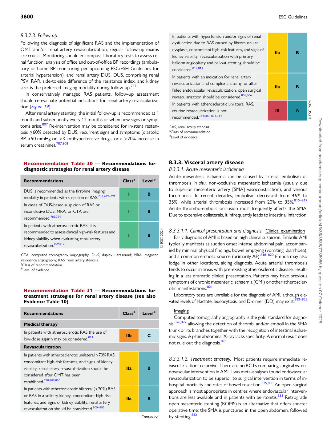
p.63 抽出テキスト
8.3.2.3 Follow-up

Following the diagnosis of significant RAS and the implementation of OMT and/or renal artery revascularization, regular follow-up exams are crucial. Monitoring should encompass laboratory tests to assess renal function, analysis of office and out-of-office BP recordings (ambulatory or home BP monitoring per upcoming ESC/ESH Guidelines for arterial hypertension), and renal artery DUS. DUS, comprising renal PSV, RAR, side-to-side difference of the resistance index, and kidney size, is the preferred imaging modality during follow-up.787 In conservatively managed RAS patients, follow-up assessment should re-evaluate potential indications for renal artery revascularization (Figure 19). After renal artery stenting, the initial follow-up is recommended at 1 month and subsequently every 12 months or when new signs or symptoms arise.807 Re-intervention may be considered for in-stent restenosis ≥60% detected by DUS, recurrent signs and symptoms (diastolic BP >90 mmHg on >3 antihypertensive drugs, or a >20% increase in serum creatinine).787,808

8.3.3 Visceral artery disease

8.3.3.1. Acute mesenteric ischaemia Acute mesenteric ischaemia can be caused by arterial embolism or thrombosis in situ, non-occlusive mesenteric ischaemia (usually due to superior mesenteric artery [SMA] vasoconstriction), and venous thrombosis. In recent decades, embolism decreased from 46% to 35%, while arterial thrombosis increased from 20% to 35%.815–817 Acute thrombo-embolic occlusion most frequently affects the SMA. Due to extensive collaterals, it infrequently leads to intestinal infarction.

8.3.3.1.1 Clinical presentation and diagnosis. Clinical examination

Early diagnosis of AMI is based on high clinical suspicion. Embolic AMI typically manifests as sudden onset intense abdominal pain, accompanied by minimal physical findings, bowel emptying (vomiting, diarrhoea), and a common embolic source (primarily AF).818–820 Emboli may also lodge in other locations, aiding diagnosis. Acute arterial thrombosis tends to occur in areas with pre-existing atherosclerotic disease, resulting in a less dramatic clinical presentation. Patients may have previous symptoms of chronic mesenteric ischaemia (CMI) or other atherosclerotic manifestations.821 Laboratory tests are unreliable for the diagnosis of AMI, although elevated levels of l-lactate, leucocytosis, and D-dimer (DD) may exist.822–825 Imaging Computed tomography angiography is the gold standard for diagnosis,826,827 allowing the detection of thrombi and/or emboli in the SMA trunk or its branches together with the recognition of intestinal ischaemic signs. A plain abdominal X-ray lacks specificity. A normal result does not rule out the diagnosis.828

8.3.3.1.2 Treatment strategy. Most patients require immediate re-

vascularization to survive. There are no RCTs comparing surgical vs. endovascular intervention in AMI. Two meta-analyses found endovascular revascularization to be superior to surgical intervention in terms of inhospital mortality and rates of bowel resection.829,830 An open surgical approach is most appropriate in centres where endovascular interventions are less available and in patients with peritonitis.831 Retrograde open mesenteric stenting (ROMS) is an alternative that offers shorter operative time; the SMA is punctured in the open abdomen, followed by stenting.832

Recommendation Table 30 — Recommendations for diagnostic strategies for renal artery disease

Recommendations Classa Levelb

DUS is recommended as the first-line imaging modality in patients with suspicion of RAS.787,789–791 I B In cases of DUS-based suspicion of RAS or inconclusive DUS, MRA, or CTA are recommended.789,791 I B In patients with atherosclerotic RAS, it is recommended to assess clinical high-risk features and kidney viability when evaluating renal artery revascularization.809,810 I B © ESC 2024

CTA, computed tomography angiography; DUS, duplex ultrasound; MRA, magnetic resonance angiography; RAS, renal artery stenosis. aClass of recommendation. bLevel of evidence.

Recommendation Table 31 — Recommendations for treatment strategies for renal artery disease (see also Evidence Table 10)

Recommendations Classa Levelb

Medical therapy In patients with atherosclerotic RAS the use of low-dose aspirin may be considered.811 IIb C Revascularization In patients with atherosclerotic unilateral >70% RAS, concomitant high-risk features, and signs of kidney viability, renal artery revascularization should be considered after OMT has been established.798,809,810 IIa B In patients with atherosclerotic bilateral (>70%) RAS or RAS in a solitary kidney, concomitant high risk features, and signs of kidney viability, renal artery revascularization should be considered.800–802 IIa B

Continued In patients with hypertension and/or signs of renal

dysfunction due to RAS caused by fibromuscular dysplasia, concomitant high-risk features, and signs of kidney viability, revascularization with primary balloon angioplasty and bailout stenting should be considered.812,813 IIa B In patients with an indication for renal artery revascularization and complex anatomy, or after failed endovascular revascularization, open surgical revascularization should be considered.805,806 IIa B In patients with atherosclerotic unilateral RAS, routine revascularization is not recommended.324,800–804,814 III A © ESC 2024

RAS, renal artery stenosis. aClass of recommendation. bLevel of evidence. Downloaded from academic.oup.com/eurheartj/article/45/36/3538/7738955 by guest on 07 October 2026