p.53 抽出テキスト
8.1.3.4 Follow-up

After revascularization or amputation, haemodynamic success should be established, aetiology of ALI investigated, and OMT ensured. Statins improve outcomes after revascularization.552,630 Since ALI is frequently caused by thrombo-embolism, Holter-ECG, echocardiogram, and aortic imaging are useful to allow initiation of appropriate therapy, in particular anticoagulation.650 Additionally, consider other prothrombotic syndromes, such as antiphospholipid syndromes and vasculitis, if clinically suspected. While there is only sparse evidence, the inclusion of PAD patients after revascularization into structured follow-up may improve their functional outcomes.627 8.2 Extracranial carotid and vertebral

artery disease 8.2.1. Clinical presentation and diagnosis 8.2.1.1. Clinical presentation Atherosclerotic CS represents one of the major causes of acute ischaemic stroke (20%).657

CS may be revealed by a cervical bruit, but also by a TIA or stroke.

8.2.1.2 Diagnosis

Atherosclerotic lesions are primarily located in specific arterial segments, including the carotid bifurcation, siphon, M1 segment of the middle cerebral artery, brachiocephalic trunk, subclavian artery, first and fourth segments of the vertebral artery, or first segment of the basilar artery. Carotid plaques (CP), originating in the intima, offer a better representation of the atherosclerotic process than carotid intima-media thickness (cIMT). CP may be diffuse or focal (protuberant). According to the Mannheim carotid plaque consensus, a CP is defined as a focal structure encroaching into the arterial lumen by ≥0.5 mm or ≥50% of the surrounding vessel.658 The American Society of Echocardiography (ASE) recently proposed a definition that includes any focal thickening considered atherosclerotic in origin and encroaching into the lumen of any carotid artery segment (protuberant-type plaque) or, in the case of diffuse vessel wall atherosclerosis, when cIMT measures ≥1.5 mm in any carotid artery segment.659 Plaques can progress to CS, defined as ≥50% narrowing of the extracranial internal carotid artery (ICA), with stenosis severity estimated using the North American Symptomatic Carotid Endarterectomy Trial (NASCET) method or its non-invasive equivalent assessed by DUS (Figure 17).122,660 Other methods are described in the Supplementary data online, Section 1.5. The European Carotid Surgery Trial (ECST) and the area methods overestimate the severity of the CS and are not recommended.77 Carotid DUS is safe, accurate, and reliable if performed by a skilled vascular specialist. It is the first-line imaging modality for screening, diagnosis, and surveillance of extracranial carotid arteries.77 The degree of stenosis is mostly based on Doppler analysis of blood flow in the common carotid artery (CCA), ICA, and external carotid artery (Table 10).661,662 Vertebral and subclavian arteries must also be checked. In some cases, indirect signs of severe stenosis have to be evaluated by transcranial and/or ophthalmic artery Doppler. Severe arterial calcification can decrease DUS accuracy.122

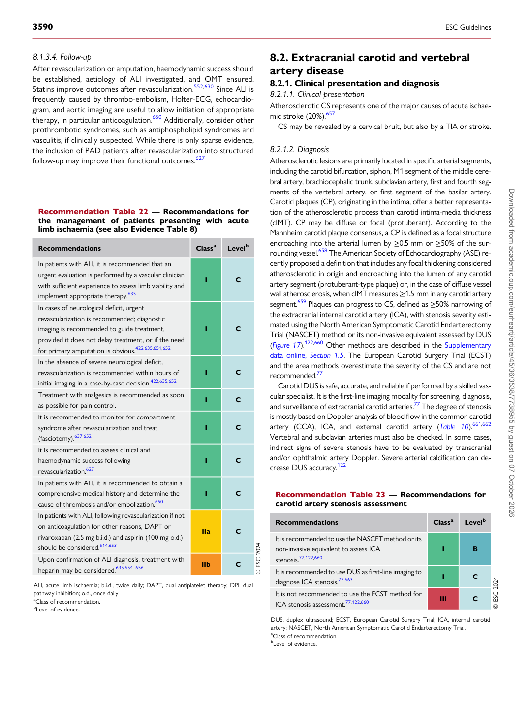
Recommendation Table 22 — Recommendations for the management of patients presenting with acute limb ischaemia (see also Evidence Table 8)

Recommendations Classa Levelb

In patients with ALI, it is recommended that an urgent evaluation is performed by a vascular clinician with sufficient experience to assess limb viability and implement appropriate therapy.635 I C In cases of neurological deficit, urgent revascularization is recommended; diagnostic imaging is recommended to guide treatment, provided it does not delay treatment, or if the need for primary amputation is obvious.422,635,651,652 I C In the absence of severe neurological deficit, revascularization is recommended within hours of initial imaging in a case-by-case decision.422,635,652 I C Treatment with analgesics is recommended as soon as possible for pain control. I C It is recommended to monitor for compartment syndrome after revascularization and treat (fasciotomy).637,652 I C It is recommended to assess clinical and haemodynamic success following revascularization.627 I C In patients with ALI, it is recommended to obtain a comprehensive medical history and determine the cause of thrombosis and/or embolization.650 I C In patients with ALI, following revascularization if not on anticoagulation for other reasons, DAPT or rivaroxaban (2.5 mg b.i.d.) and aspirin (100 mg o.d.) should be considered.514,653 IIa C Upon confirmation of ALI diagnosis, treatment with heparin may be considered.635,654–656 IIb C © ESC 2024

ALI, acute limb ischaemia; b.i.d., twice daily; DAPT, dual antiplatelet therapy; DPI, dual pathway inhibition; o.d., once daily. aClass of recommendation. bLevel of evidence. Recommendation Table 23 — Recommendations for carotid artery stenosis assessment

Recommendations Classa Levelb

It is recommended to use the NASCET method or its non-invasive equivalent to assess ICA stenosis.77,122,660 I B It is recommended to use DUS as first-line imaging to diagnose ICA stenosis.77,663 I C It is not recommended to use the ECST method for ICA stenosis assessment.77,122,660 III C © ESC 2024

DUS, duplex ultrasound; ECST, European Carotid Surgery Trial; ICA, internal carotid artery; NASCET, North American Symptomatic Carotid Endarterectomy Trial. aClass of recommendation. bLevel of evidence. Downloaded from academic.oup.com/eurheartj/article/45/36/3538/7738955 by guest on 07 October 2026