p.51 抽出テキスト
8.1.3.1.1 Clinical examination. The emergency level and the choice

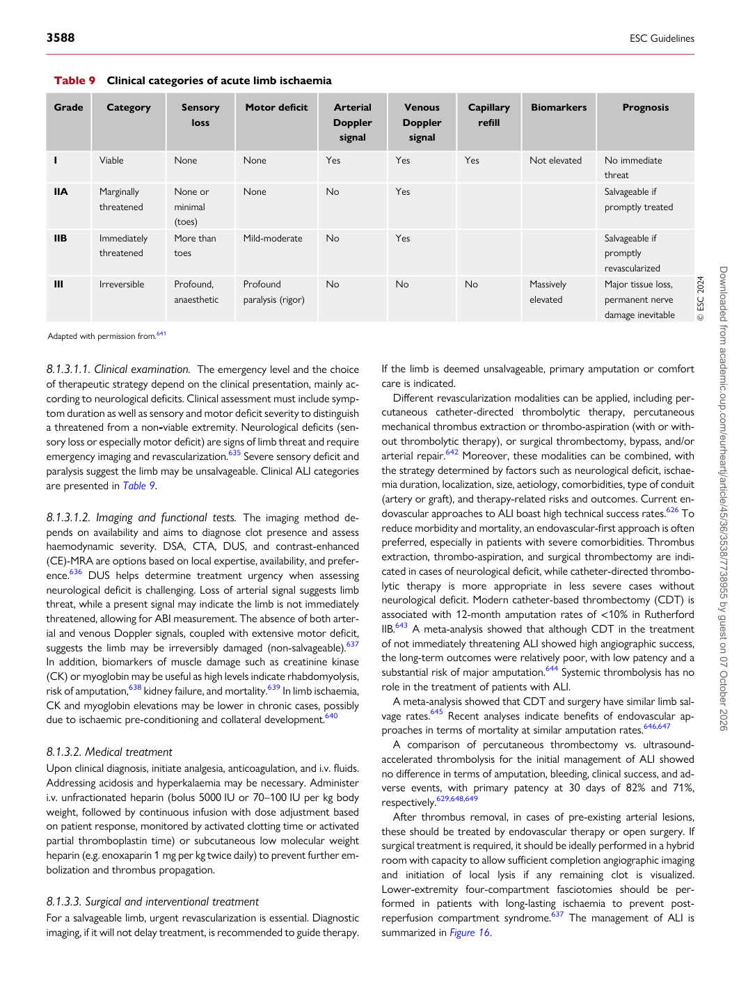
of therapeutic strategy depend on the clinical presentation, mainly according to neurological deficits. Clinical assessment must include symptom duration as well as sensory and motor deficit severity to distinguish a threatened from a non-viable extremity. Neurological deficits (sensory loss or especially motor deficit) are signs of limb threat and require emergency imaging and revascularization.635 Severe sensory deficit and paralysis suggest the limb may be unsalvageable. Clinical ALI categories are presented in Table 9.

8.1.3.1.2 Imaging and functional tests. The imaging method de-

pends on availability and aims to diagnose clot presence and assess haemodynamic severity. DSA, CTA, DUS, and contrast-enhanced (CE)-MRA are options based on local expertise, availability, and preference.636 DUS helps determine treatment urgency when assessing neurological deficit is challenging. Loss of arterial signal suggests limb threat, while a present signal may indicate the limb is not immediately threatened, allowing for ABI measurement. The absence of both arterial and venous Doppler signals, coupled with extensive motor deficit, suggests the limb may be irreversibly damaged (non-salvageable).637 In addition, biomarkers of muscle damage such as creatinine kinase (CK) or myoglobin may be useful as high levels indicate rhabdomyolysis, risk of amputation,638 kidney failure, and mortality.639 In limb ischaemia, CK and myoglobin elevations may be lower in chronic cases, possibly due to ischaemic pre-conditioning and collateral development.640

8.1.3.2 Medical treatment

Upon clinical diagnosis, initiate analgesia, anticoagulation, and i.v. fluids. Addressing acidosis and hyperkalaemia may be necessary. Administer i.v. unfractionated heparin (bolus 5000 IU or 70–100 IU per kg body weight, followed by continuous infusion with dose adjustment based on patient response, monitored by activated clotting time or activated partial thromboplastin time) or subcutaneous low molecular weight heparin (e.g. enoxaparin 1 mg per kg twice daily) to prevent further embolization and thrombus propagation.

8.1.3.3 Surgical and interventional treatment

For a salvageable limb, urgent revascularization is essential. Diagnostic imaging, if it will not delay treatment, is recommended to guide therapy. If the limb is deemed unsalvageable, primary amputation or comfort care is indicated.

Different revascularization modalities can be applied, including percutaneous catheter-directed thrombolytic therapy, percutaneous mechanical thrombus extraction or thrombo-aspiration (with or without thrombolytic therapy), or surgical thrombectomy, bypass, and/or arterial repair.642 Moreover, these modalities can be combined, with the strategy determined by factors such as neurological deficit, ischaemia duration, localization, size, aetiology, comorbidities, type of conduit (artery or graft), and therapy-related risks and outcomes. Current endovascular approaches to ALI boast high technical success rates.626 To reduce morbidity and mortality, an endovascular-first approach is often preferred, especially in patients with severe comorbidities. Thrombus extraction, thrombo-aspiration, and surgical thrombectomy are indicated in cases of neurological deficit, while catheter-directed thrombolytic therapy is more appropriate in less severe cases without neurological deficit. Modern catheter-based thrombectomy (CDT) is associated with 12-month amputation rates of <10% in Rutherford IIB.643 A meta-analysis showed that although CDT in the treatment of not immediately threatening ALI showed high angiographic success, the long-term outcomes were relatively poor, with low patency and a substantial risk of major amputation.644 Systemic thrombolysis has no role in the treatment of patients with ALI. A meta-analysis showed that CDT and surgery have similar limb salvage rates.645 Recent analyses indicate benefits of endovascular approaches in terms of mortality at similar amputation rates.646,647 A comparison of percutaneous thrombectomy vs. ultrasoundaccelerated thrombolysis for the initial management of ALI showed no difference in terms of amputation, bleeding, clinical success, and adverse events, with primary patency at 30 days of 82% and 71%, respectively.629,648,649 After thrombus removal, in cases of pre-existing arterial lesions, these should be treated by endovascular therapy or open surgery. If surgical treatment is required, it should be ideally performed in a hybrid room with capacity to allow sufficient completion angiographic imaging and initiation of local lysis if any remaining clot is visualized. Lower-extremity four-compartment fasciotomies should be performed in patients with long-lasting ischaemia to prevent postreperfusion compartment syndrome.637 The management of ALI is summarized in Figure 16.

Table 9 Clinical categories of acute limb ischaemia

Grade Category Sensory

loss Motor deficit Arterial

Doppler

signal Venous

Doppler

signal Capillary

refill Biomarkers Prognosis

I Viable None None Yes Yes Yes Not elevated No immediate

threat

IIA Marginally

threatened None or

minimal (toes) None No Yes Salvageable if

promptly treated

IIB Immediately threatened More than toes Mild-moderate No Yes Salvageable if promptly

revascularized

III Irreversible Profound,

anaesthetic Profound

paralysis (rigor) No No No Massively

elevated Major tissue loss,

permanent nerve damage inevitable © ESC 2024

Adapted with permission from.641 Downloaded from academic.oup.com/eurheartj/article/45/36/3538/7738955 by guest on 07 October 2026