p.48 抽出テキスト
8.1.1.3 Follow-up

Asymptomatic and symptomatic PAD are at increased risk of leg symptom worsening427 and of CV mortality and morbidity.419,431,551 Follow-up post-revascularization is crucial to ensure perfusion improvement, address CVRFs, optimize pharmacological treatment adherence, identify disease progression, and evaluate mental health and functional capacity. Experienced vascular care physicians should conduct follow-up, although specific protocols are currently undefined.128,552 Data on asymptomatic PAD follow-up are limited.553 For symptomatic PAD or postintervention, annual follow-up are advised, including ABI/TBI measurement and DUS for new or worsening symptoms.

8.1.2 Chronic limb-threatening ischaemia

8.1.2.1. Clinical presentation and diagnosis Chronic limb-thretening ischaemia describes chronic lower-limb hypoperfusion responsible for ischaemic rest pain, or non-healing ulceration or gangrene (typically in distal segments).555,556 Ischaemic rest pain primarily affects the patient’s forefoot and aggravates in a supine position, while lowering of the affected leg eases ischaemic symptoms.

8.1.2.1.1 Definition. Chronic limb-thretening ischaemia should be

considered in the presence of one of the following lower-limb clinical signs or symptoms: • Ischaemic rest pain • Non-healing lower-limb wound of ≥2 weeks’ duration • Lower-limb gangrene

The following haemodynamic criteria may be used to guide diagnosis in patients with suspicion of CLTI: • Ankle pressure <50 mmHg • TP <30 mmHg • TcPO2 <30 mmHg

8.1.2.1.2 Initial assessment and risk of amputation. For patients

with CLTI, initial diagnostic steps involve clinical examination and limb perfusion assessment through haemodynamic measurements. Regarding haemodynamic assessment in CLTI, standard ABI may be normal or falsely elevated due to non-compressible arteries related to medial sclerosis (common in diabetes or CKD),557 which can be overcome by estimation of ABI based on Doppler waveforms.124 Therefore, standard ankle pressure alone may not be reliable in estimating limb loss risk.441,558 In addition, a large proportion of patients with ulcers may have below-the-ankle lesions.440 In patients with CLTI, TP, TBI, or TcPO2 should additionally be obtained.90,441,559 Particularly in patients with CLTI, the WIfI classification system should be applied. In addition to patients’ limb perfusion, the WIfI classification considers the wound size and the extent of foot infection to determine the individual risk of amputation.417,444–446

8.1.2.1.3 Imaging. In all patients with CLTI, comprehensive vascular

imaging is mandatory to evaluate revascularization options. CLTI commonly affects more than one arterial segment of the lower limbs, involving infra-popliteal arteries (BTK and below-the-ankle arteries) in most cases. While non-invasive imaging (DUS, CTA, MRA) provides reliable results for above-the-knee arteries, imaging of BTK arteries, especially below the ankle, may be hampered by severe calcification.448,560,561 Therefore, in CLTI additional DSA with dedicated views of the foot should be considered for the assessment of BTK arteries.560 Even in patients who are not candidates for revascularization, DSA should be obtained to prevent unnecessary amputation or to minimize amputation extent.560,562

8.1.2.1.4 Mortality risk assessment. All-cause mortality and event

rates of MI are more than two-fold higher in CLTI patients than in unselected patients with an ABI ≤0.90.431 In CLTI patients undergoing revascularization, the post-revascularization period is particularly associated with an increased risk of MALE and MACE.563 The management of patients with CLTI should therefore include an individual peri-procedural risk assessment. Referring to the periprocedural risk patients can be categorized as average procedural risk (peri-procedural mortality <5% and 2 year survival >50%) or high procedural risk (peri-procedural mortality ≥5% and 2 year survival ≤50%).564,565 Besides revascularization, it also needs to be considered that lowerlimb amputation is associated with 30 day mortality rates of up to 22%.566

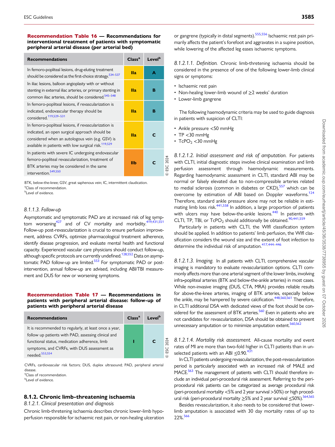
Recommendation Table 16 — Recommendations for interventional treatment of patients with symptomatic peripheral arterial disease (per arterial bed)

Recommendations Classa Levelb

In femoro-popliteal lesions, drug-eluting treatment should be considered as the first-choice strategy.534–537 IIa A In iliac lesions, balloon angioplasty with or without stenting in external iliac arteries, or primary stenting in common iliac arteries, should be considered.545–548 IIa B In femoro-popliteal lesions, if revascularization is indicated, endovascular therapy should be considered.119,529–531 IIa B In femoro-popliteal lesions, if revascularization is indicated, an open surgical approach should be considered when an autologous vein (e.g. GSV) is available in patients with low surgical risk.119,529 IIa C In patients with severe IC undergoing endovascular femoro-popliteal revascularization, treatment of BTK arteries may be considered in the same intervention.549,550 IIb C © ESC 2024

BTK, below-the-knee; GSV, great saphenous vein; IC, intermittent claudication. aClass of recommendation. bLevel of evidence.

Recommendation Table 17 — Recommendations in patients with peripheral arterial disease: follow-up of patients with peripheral arterial disease

Recommendations Classa Levelb

It is recommended to regularly, at least once a year, follow up patients with PAD, assessing clinical and functional status, medication adherence, limb symptoms, and CVRFs, with DUS assessment as needed.553,554 I C © ESC 2024

CVRFs, cardiovascular risk factors; DUS, duplex ultrasound; PAD, peripheral arterial disease. aClass of recommendation. bLevel of evidence.

ESC Guidelines                                                                                                                                                                                          3585

Downloaded from academic.oup.com/eurheartj/article/45/36/3538/7738955 by guest on 07 October 2026