p.30 抽出テキスト
(inflammation/infection) and treatment response.187,188 Although different tracers have been tested to identify calcification, fibrosis and/or thrombus formation, most PET studies have focused on vasculitis. The relationship between FDG-PET images and AAA progression is controversial. However, fluorine-18–sodium fluoride (18F–NaF) PET-computed tomography (PET-CT), a marker of active vascular calcification and high-risk plaques, has shown a correlation between increased tracer uptake, AAA growth, and CV events.189 PET-CT has shown better diagnostic accuracy in identifying lesions and detecting graft infection or infectious aortic diseases.190–193 High radiation exposure, high costs, and limited availability are the main limitations of PET.

5.4.9 Intravascular ultrasound

Intravascular ultrasound (IVUS) provides high-resolution imaging for artery and vein diseases, aiding complex aortic disease management by distinguishing true and false lumens and guiding stent placement. It is operator-dependent, costly, and less accessible, but seems to provide better measurements for acute aortic syndromes.194

5.4.10 Digital subtraction aortography

Non-invasive imaging modalities have replaced DSA in first-line diagnostic testing, both in suspected AAS or known chronic AD; however, DSA might be useful if findings in non-invasive techniques are ambiguous or incomplete. It is primarily used for the percutaneous treatment of CAD, aortic visceral branches, or for monitoring thoracic endovascular aortic aneurysm repair (TEVAR/EVAR) implantation.

6. Screening for carotid, peripheral arterial, and aortic diseases

6.1 Screening for carotid and peripheral

arterial diseases 6.1.1. Lower-extremity peripheral arterial disease Due to elevated CV risk in chronic PAD, early diagnosis, prevention, and robust cardiovascular risk factor (CVRF) control are essential, even in asymptomatic cases. ‘Intermediary CV risk’ individuals may be reclassified as ‘high or very high risk’, prompting adapted prevention. ABI is the preferred first-line test for asymptomatic individuals aged ≥65 years,14,195 especially women.196 Screening might also be beneficial at a younger age in case of CVRFs, but data are still lacking. Clinical examination, functional status, and walking capacity assessment are recommended to detect ‘masked PAD’.77 In diabetes, early PAD (and foot neuropathy) diagnosis is crucial. Effective CVRF management and treatment can prevent CV disease, foot wounds, and amputation.197 In patients with diabetes and normal resting ABI, TBI measurement should be considered. The prevalence of popliteal aneurysms (PAs) is high in patients with AAA and subaneurysmal aortic dilatation, warranting screening. PAs are correlated with iliac and femoral artery diameters.76 In patients needing transfemoral access, screening for iliofemoral artery disease may be considered.198 6.1.2 Carotid artery stenosis

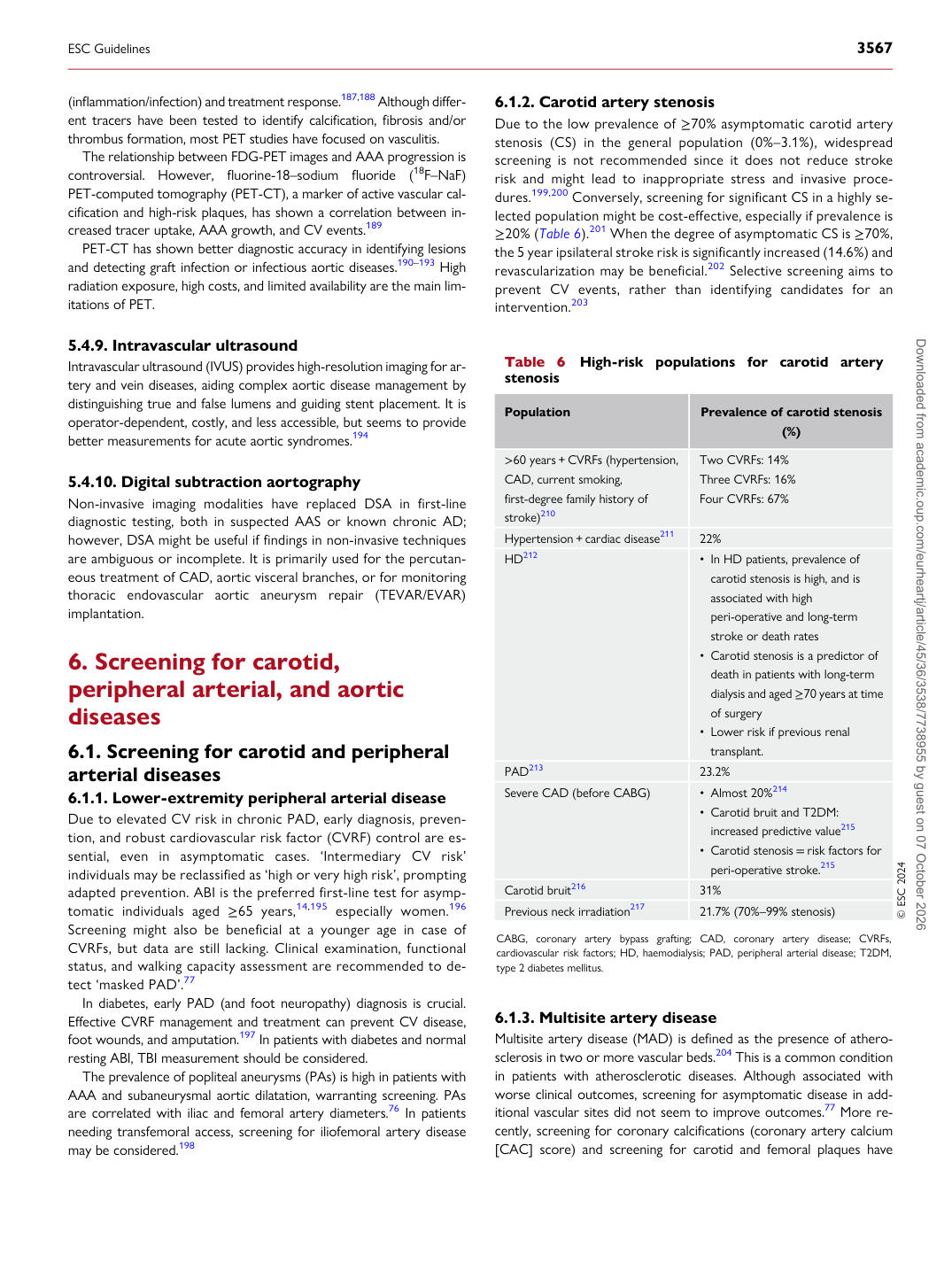
Due to the low prevalence of ≥70% asymptomatic carotid artery stenosis (CS) in the general population (0%–3.1%), widespread screening is not recommended since it does not reduce stroke risk and might lead to inappropriate stress and invasive procedures.199,200 Conversely, screening for significant CS in a highly selected population might be cost-effective, especially if prevalence is ≥20% (Table 6).201 When the degree of asymptomatic CS is ≥70%, the 5 year ipsilateral stroke risk is significantly increased (14.6%) and revascularization may be beneficial.202 Selective screening aims to prevent CV events, rather than identifying candidates for an intervention.203

6.1.3 Multisite artery disease

Multisite artery disease (MAD) is defined as the presence of atherosclerosis in two or more vascular beds.204 This is a common condition in patients with atherosclerotic diseases. Although associated with worse clinical outcomes, screening for asymptomatic disease in additional vascular sites did not seem to improve outcomes.77 More recently, screening for coronary calcifications (coronary artery calcium [CAC] score) and screening for carotid and femoral plaques have

Table 6 High-risk populations for carotid artery stenosis

Population Prevalence of carotid stenosis

(%)

>60 years + CVRFs (hypertension, CAD, current smoking, first-degree family history of stroke)210 Two CVRFs: 14%

Three CVRFs: 16% Four CVRFs: 67%

Hypertension + cardiac disease211 22% HD212 • In HD patients, prevalence of

carotid stenosis is high, and is associated with high peri-operative and long-term stroke or death rates • Carotid stenosis is a predictor of

death in patients with long-term dialysis and aged ≥70 years at time of surgery • Lower risk if previous renal

transplant.

PAD213 23.2% Severe CAD (before CABG) • Almost 20%214

• Carotid bruit and T2DM:

increased predictive value215 • Carotid stenosis = risk factors for

peri-operative stroke.215

Carotid bruit216 31% Previous neck irradiation217 21.7% (70%–99% stenosis) © ESC 2024

CABG, coronary artery bypass grafting; CAD, coronary artery disease; CVRFs, cardiovascular risk factors; HD, haemodialysis; PAD, peripheral arterial disease; T2DM, type 2 diabetes mellitus.

ESC Guidelines                                                                                                                                                                                          3567

Downloaded from academic.oup.com/eurheartj/article/45/36/3538/7738955 by guest on 07 October 2026