p.29 抽出テキスト
Transoesophageal echocardiography (TOE) is highly accurate (sensitivity: up to 99%, specificity: 89% for AAS), except with absolute contraindications like oesophageal issues, bleeding, recent gastro-oesophageal surgery, or respiratory distress. TOE is convenient for bedside and intraoperative use but less suitable for long-term surveillance, which requires evaluation with CCT/CMR.

5.4.5 Duplex ultrasound imaging of the abdominal

aorta After scanning both transversally and longitudinally, the anteroposterior (AP) diameter in a cross-sectional view of the AA should be measured. Ensure the DUS beam is perpendicular to the AA axis, forming a circular vessel section. If the AA is sinuous or dilated, achieving equal AP and transverse diameters may be challenging. In such instances, calculate the mean ellipse diameter or measure the AA diameter in a clear longitudinal view with a perpendicular diameter (Figure 6).122 The outer-to-outer (Figure 6) method is the one recommended by the American Institute of Ultrasound in Medicine, the American College of Cardiology/American Heart Association (ACC/AHA), and the European Society of Cardiology (ESC), since it is more reliable in cases of atherosclerotic plaque or intravascular thrombus and best correlates with CCT and CMR. However, the most effective methodology is under debate and further studies are needed to determine the best convention.169

Normal diameters of the AA are reported in Figure 5 and Supplementary data online, Section 1.3.

5.4.6 Cardiovascular computed tomography

Cardiovascular computed tomography, due to its quick acquisition, wide availability, high reproducibility, and suitability for emergency departments, is the primary imaging method for aortic disease diagnosis, prognosis, and therapy planning (sensitivity 100%, specificity 98% for AAS).170–172 ‘Double or triple rule-out’ protocols concurrently assess the aorta, pulmonary, and coronary arteries.173,174 Electrocardiogram triggering is crucial to prevent motion artefacts (especially in the aortic root and ascending aorta), which can distort measurements or resemble dissection flaps, facilitating coronary artery assessment. The standard protocol comprises non-enhanced scans (for calcification, IMH, or surgical material), contrast-enhanced CCT angiography, and a late scan (to visualize contrast leakage or aortic wall late enhancement suggestive of inflammation or infection).175 Iodinated contrast agents carry potential allergic reactions and postcontrast acute kidney injury (PC-AKI) risks.176 In these cases, opt for contrast-free CCT for accurate aortic diameter measurement (also for CMR-intolerant patients). Moreover, excessive radiation caution is crucial, particularly in young females, when performing CCT for monitoring chronic aortic diseases.177

5.4.7 Cardiovascular magnetic resonance

Cardiovascular magnetic resonance comprehensively evaluates the aorta, including shape, diameter, tissue characteristics (inflammation, infection, atheroma, bleeding),178 lesion extent, side branches, adjacent structures, and mural thrombus. It assesses ventricular and valve function, quantifies flow, and employs cine steady-state free precession (SSFP) or ECG-gated angio-CMR for the aortic root, while non-gated sequences suffice for the rest. Recently, 4D flow sequences179 have been developed to evaluate complex intravascular flows,180,181 complex flow parameters (wall shear stress, pulse wave velocity, or kinetic energy), or flow quantification at different levels in one unique acquisition (useful in AD or congenital diseases).182,183 Cardiovascular magnetic resonance obviates ionizing radiation and iodinated contrast (3D contrast CMR), making it ideal for young patients, women, and pregnancy. Caution is warranted, especially with non-macrocyclic gadolinium, for estimated glomerular filtration rate (eGFR) <30 mL/min/1.73 m2 (Supplementary data online, Section 1.2). CMR is increasingly used in patients with intracardiac devices (pacemakers/implantable cardioverter defibrillators, CMR- and non-CMR-compatible devices) with proper monitoring, but not for those with cochlear implants or intracranial clips.184,185 In the acute setting, CMR use is limited because of low availability, difficulties in monitoring unstable patients, and longer acquisition times.166,186

5.4.8 Positron emission tomography

Positron emission tomography (PET) usually uses 18F-fluorodeoxyglucose (FDG), allowing non-invasive assessments of metabolic activity

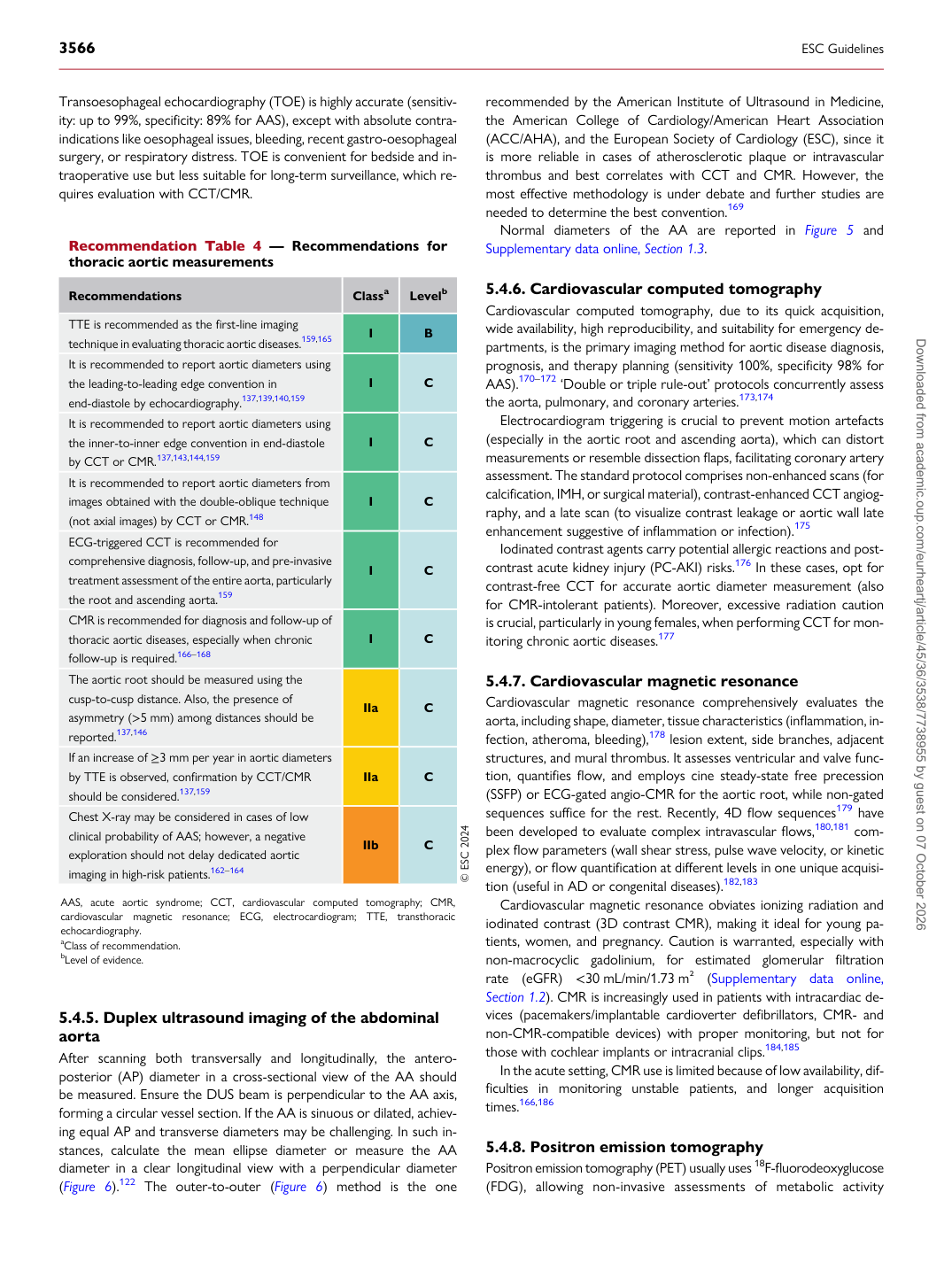
Recommendation Table 4 — Recommendations for thoracic aortic measurements

Recommendations Classa Levelb

TTE is recommended as the first-line imaging technique in evaluating thoracic aortic diseases.159,165 I B It is recommended to report aortic diameters using the leading-to-leading edge convention in end-diastole by echocardiography.137,139,140,159 I C It is recommended to report aortic diameters using the inner-to-inner edge convention in end-diastole by CCT or CMR.137,143,144,159 I C It is recommended to report aortic diameters from images obtained with the double-oblique technique (not axial images) by CCT or CMR.148 I C ECG-triggered CCT is recommended for comprehensive diagnosis, follow-up, and pre-invasive treatment assessment of the entire aorta, particularly the root and ascending aorta.159 I C CMR is recommended for diagnosis and follow-up of thoracic aortic diseases, especially when chronic follow-up is required.166–168 I C The aortic root should be measured using the cusp-to-cusp distance. Also, the presence of asymmetry (>5 mm) among distances should be reported.137,146 IIa C If an increase of ≥3 mm per year in aortic diameters by TTE is observed, confirmation by CCT/CMR should be considered.137,159 IIa C Chest X-ray may be considered in cases of low clinical probability of AAS; however, a negative exploration should not delay dedicated aortic imaging in high-risk patients.162–164 IIb C © ESC 2024

AAS, acute aortic syndrome; CCT, cardiovascular computed tomography; CMR, cardiovascular magnetic resonance; ECG, electrocardiogram; TTE, transthoracic echocardiography. aClass of recommendation. bLevel of evidence. Downloaded from academic.oup.com/eurheartj/article/45/36/3538/7738955 by guest on 07 October 2026