p.28 抽出テキスト
Given the high incidence of atherosclerotic plaques/thrombi in the AA, the outer-to-outer convention should be preferred (also presenting the best agreement with CCT and CMR) (Figure 6).141,142 Regarding CCT and CMR, measurements must be performed using the inner-to-inner edge method (Figure 6) in end-diastole (fewer motion artefacts).137,143,144 The aortic root is measured in the parasternal long axis by transthoracic echocardiography (TTE),137,139,140,145 since the short axis underestimates the diameter due to possible plane obliquity. By CMR or CCT, the cusp-to-cusp diameter best correlates with echocardiography (Figure 6). A diameter difference >5 mm (among root diameters within the same imaging modality) indicates root asymmetry, frequent in bicuspid aortic valve (BAV) or genetic aortopathies, which is important to be determined since it generates underestimations.146 While 3D echocardiography is a potential surveillance alternative in these cases (especially if CMR/CCT is limited for serial follow-up), validation studies are lacking.147 In end-diastole, measure the ascending aorta by moving the transducer 1–2 intercostal spaces up in the parasternal long axis. Echocardiography provides information on aortic arch or DTA enlargement, but diagnostic certainty (precise measurement of the diameters) is lacking. CCT or CMR uses the double-oblique technique to measure aortic diameters, reporting antero-posterior and perpendicular dimensions for accurate assessment.148 It is recommended to report aortic measurements by specific segments based on anatomical landmarks and to relate the largest diameter to a nearby anatomical structure for reference. Changes in aortic diameter require a ≥3 mm increase in echocardiography, which should be confirmed with CCT/CMR and compared with baseline measurements. For accurate assessment, stick to the same imaging technique, centre, methodology, and side-by-side comparisons.137,140

5.4.2 Normal aortic values

When evaluating aortic dimensions and clinical relevance, consider factors like aortic region, anthropometric measurements, patient history, and underlying medical conditions. Factors influencing aortic and peripheral artery size in the normal population include age, sex, ethnicity, body surface area (BSA), and, particularly, height.149 Body surface area is the most used method to normalize aortic dimensions based on an individual’s body size, thus an ascending thoracic aorta >22 mm/m2 or a DTA >16 mm/m2 is considered aortic dilatation.150–152 However, extremes of low or high body weight pose limitations. In such cases, surgical thresholds may involve indexing aortic diameter by height (an aorta height index >32.1 mm/m is associated with a 12% yearly risk of aortic adverse events [AAE]),153 aortic cross-sectional area to patient height (a ratio ≥10 cm2/m implies reduced long-term survival),154 or aortic length (from the aortic annulus to the innominate artery, considering a length >11 cm a threshold for surgery).155 To correlate measured diameter with the expected one based on age, sex, and body surface, use nomograms or z-score calculation formulas, especially in heritable thoracic aortic disease (HTAD). Supplementary data online, Figure S1 and Table S2, presents nomograms developed for echocardiography, applicable also to CCT and CMR.156,157 Calculation of z-scores can be performed following these links: https://www.marfan.fr/accueil/z-score-calculus/ or https:// marfan.org/dx/z-score-adults/; reference values used for their estimation may vary depending on age and other factors. However, z-scores are limited by the fact that not all ethnic groups are equally represented (mostly white) and over- or underweight can lead to an over- or underestimation.158 Moreover, with ageing and loss of elastic properties, the aorta tends to enlarge. Aortic growth in adults is about 0.9 mm per 10 years in males and 0.7 mm per 10 years in females, which may be influenced by BP, physical activity, and genetic factors.

5.4.3 Chest X-ray and electrocardiogram

Chest X-ray obtained for other indications in asymptomatic patients or in cases of acute aortic syndrome (AAS) suspicion may detect abnormalities of aortic size/contour that need to be confirmed by another imaging technique. It presents limited sensitivity (64%) and specificity (86%) in the diagnosis of aortic diseases;162 thus, a normal chest X-ray may not rule out the diagnosis of AAS.162–164 On the contrary, chest X-ray may identify other causes of chest pain (e.g. pleural effusion or pneumothorax). Electrocardiogram (ECG) might be useful to rule out other causes of chest pain (e.g. MI) or AAS complications (coronary occlusion/dissection) but it is not useful for AAS diagnosis.

5.4.4 Echocardiography

It is considered the first-line imaging technique in the evaluation of aortic disease, assessing all echocardiographic windows and the aortic valve. It provides key anatomic information (i.e. dilatation, atherosclerotic lesions, or dissection) for the ascending aorta, arch, and AA; however, it is not useful to assess the exact diameters of the aortic arch and DTA (requiring confirmation with CCT/CMR). Also, the distal ascending aorta and proximal arch (blind spot) are inadequately visualized due to left mainstem bronchus interposition. Transthoracic echocardiography can identify AAS complications (e.g. aortic regurgitation, tamponade, or wall motion abnormalities), but its diagnostic accuracy for AAS is limited (sensitivity: 78%–100% for type A, 31%–55% for type B). Contrast enhancement improves diagnosis.165

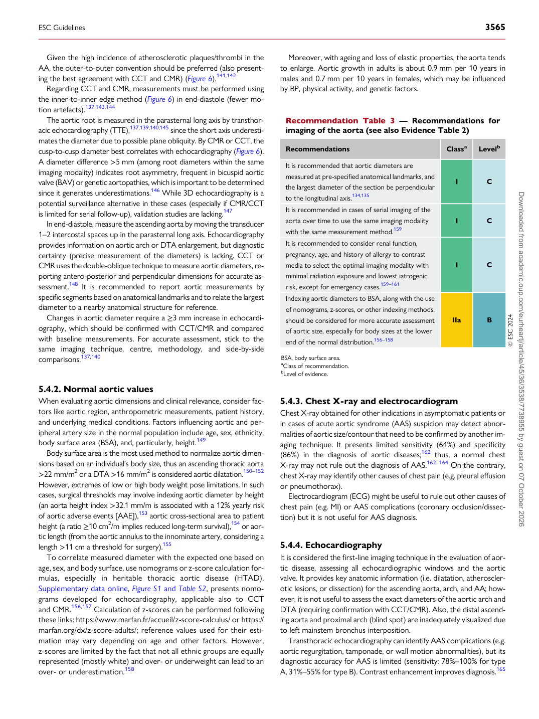
Recommendation Table 3 — Recommendations for imaging of the aorta (see also Evidence Table 2)

Recommendations Classa Levelb

It is recommended that aortic diameters are measured at pre-specified anatomical landmarks, and the largest diameter of the section be perpendicular to the longitudinal axis.134,135 I C It is recommended in cases of serial imaging of the aorta over time to use the same imaging modality with the same measurement method.159 I C It is recommended to consider renal function, pregnancy, age, and history of allergy to contrast media to select the optimal imaging modality with minimal radiation exposure and lowest iatrogenic risk, except for emergency cases.159–161 I C Indexing aortic diameters to BSA, along with the use of nomograms, z-scores, or other indexing methods, should be considered for more accurate assessment of aortic size, especially for body sizes at the lower end of the normal distribution.156–158 IIa B © ESC 2024

BSA, body surface area. aClass of recommendation. bLevel of evidence.

ESC Guidelines                                                                                                                                                                                          3565

Downloaded from academic.oup.com/eurheartj/article/45/36/3538/7738955 by guest on 07 October 2026