p.25 抽出テキスト
range from 45% to 100% and 17% to 100%, respectively.91 The usual pathological threshold for TBI is ≤0.70 (Figure 4).99 Used within the Framingham risk score, ABI enables the upgrading of risk estimation in ‘low-risk’ women and men,77,88 it allows CV risk assessment in diverse ethnic groups independently of risk factors,77,89 and is inexpensive and minimally time-consuming.100 Trained physicians have better reproducibility than inexperienced ones.101,102 In patients with exertional limb pain relieved by rest and a resting ABI >0.90, exercise testing with post-exercise ABI measurements or exercise oximetry has been proposed to diagnose lower-limb arterial stenoses.103–105 The post-exercise ABI is determined 1 min after the cessation of a standardized treadmill exercise.106 The physician measures bilateral ankle BP, starting with the symptomatic leg, using the ankle artery used for the reference resting ABI measurement. Brachial SBP should simultaneously be measured to enable calculation of the post-exercise ABI.104 Discrepancies in PAD diagnosis exist between exercise criteria, such as a fall in absolute ankle BP >30 mmHg or a drop of >20% in the postexercise ABI.104 Recent studies identified numerous false positives in a healthy population when using a post-exercise ABI drop of >20% as the diagnostic threshold, as commonly proposed.103 Measurement of transcutaneous oxygen pressure (TcPO2) is a means of evaluating tissue viability and is proposed as a diagnostic criterion of CLTI (Figure 4).107 TcPO2 is affected by local and general factors such as skin thickness, probe temperature, inflammation, and oedema,108,109 resulting in misleading values. Resting TcPO2 >30 mmHg is a favourable indicator of wound healing;110–112 however, resting TcPO2 <10 mmHg is associated with bad prognosis for wound healing and amputation in CLTI patients treated with bone marrow-derived stem cells.107 When performed at successive levels on an ischaemic limb, TcPO2 measurement may help to determine amputation level.113–115

Exercise transcutaneous oximetry has also been proposed.116,117 This seems of interest to detect proximal (buttock) claudication105 or unsuspected exercise-induced hypoxaemia118 in patients with intermittent claudication (IC).117

5.3.1 Duplex ultrasound

Duplex ultrasound (DUS) is a first step in the vascular work-up for PAD screening and diagnosis, allowing a dynamic, non-invasive, radiation- and contrast-free examination. It localizes vascular lesions and quantifies their extent and severity through velocity criteria.119–121 In combination with ABI or TBI, DUS permits determining the haemodynamic relevance of arterial lesions122,123 and estimation of ABI.124 DUS has a sensitivity of 88% and specificity of 95% for >50% stenosis detection.125 Post-exercise DUS can reveal borderline arterial lesions if initial findings are inconclusive.122,126,127 Duplex ultrasound distinguishes atherosclerotic (even subclinical disease) from non-atherosclerotic lesions, but its reliability relies on the sonographer’s expertise.122 Cross-sectional imaging is advisable for revascularization planning. ABI and DUS are recommended for PAD patient follow-up post-revascularization.128 More recent techniques, such as flow imaging, 3D echography, ultrafast ultrasound, and shear wave elastography, as well as the use of contrast-enhanced ultrasound (CEUS), could further improve DUS performance.129 5.3.2 Digital subtraction angiography, computed

tomography angiography, and magnetic resonance angiography Detailed information about these techniques can be found in the Supplementary data online, Section 1.2 (Table S1). Digital subtraction angiography (DSA) remains mostly limited to revascularization procedures. Computed tomography angiography (CTA) offers better spatial resolution than magnetic resonance angiography (MRA) and better calcification visualization; however, it can also overestimate stenosis severity due to the blooming effect. MRA allows arterial wall and lumen assessment as well as tissue and organ perfusion distal to or surrounding the explored arterial territory.

5.4 Evaluation of the aorta

The aorta can be divided into different anatomical regions (from proximal to distal) for reporting purposes. The main anatomical aortic regions are the aortic root, ascending aorta, aortic arch, descending thoracic aorta (DTA), abdominal aorta (AA), infrarenal aorta, and the iliac arteries (Figure 5).134,135

5.4.1 Aortic measurements

The main imaging techniques used for aortic evaluation are illustrated in Table 5. Evaluating aortic dilation and progression depends on standardized measurements. In echocardiography, aortic diameters should be measured using the leading-to-leading edge method during end-diastole (as systole sees about a 2 mm aortic expansion) in all segments (Figure 6).137,138 Most studies supporting prophylactic surgery have used this approach. Furthermore, better agreement exists between echo’s leading-to-leading edge and cardiovascular computed tomography (CCT)/cardiovascular magnetic resonance (CMR)’s inner-to-inner edge during end-diastole.137,139,140 However, when the aortic wall thickens (e.g. atheroma, thrombus, intramural haematoma [IMH], or aortitis) or in cases of aortic dissection (AD), also report the outer-to-outer diameter (Figure 6).

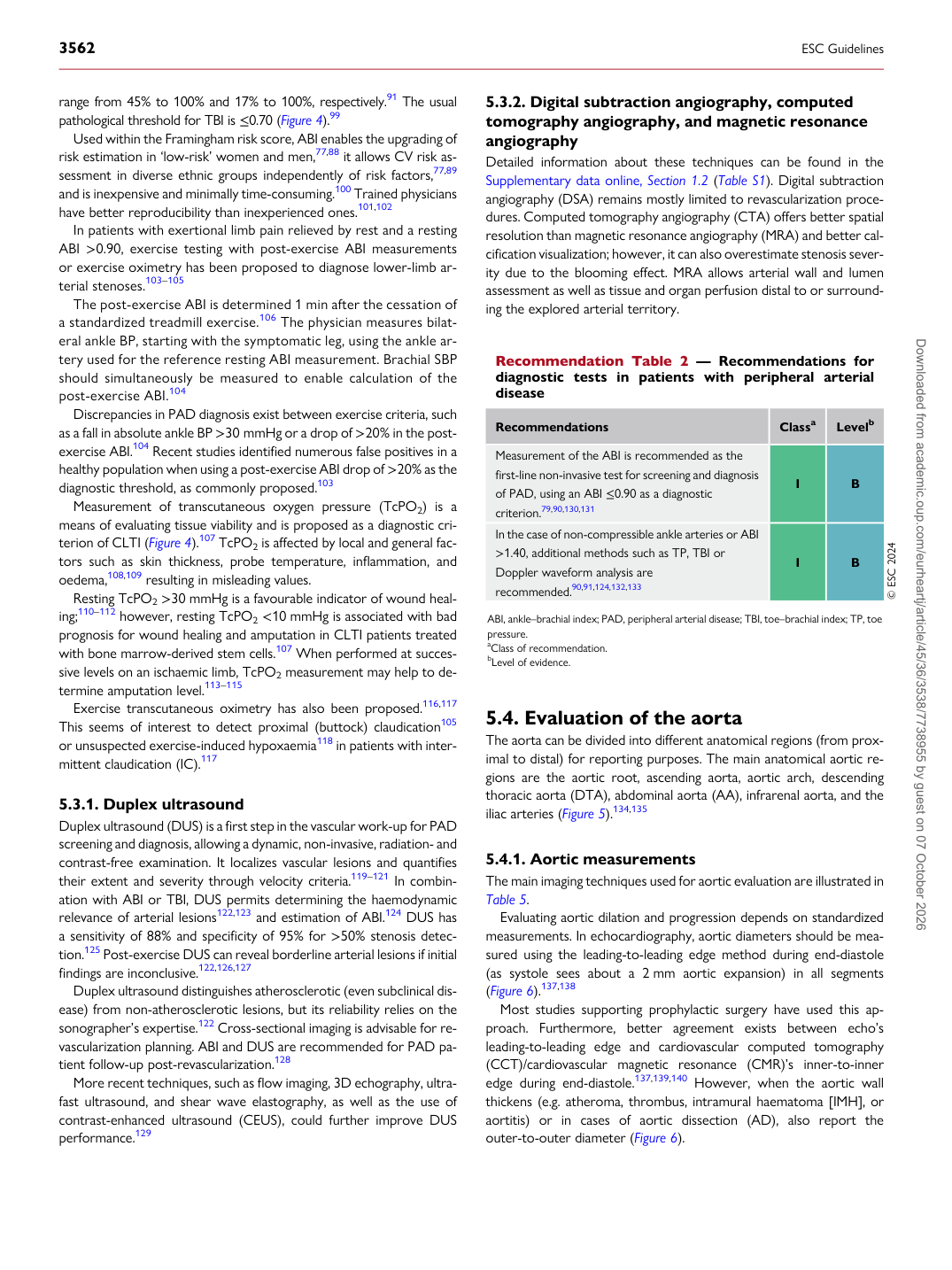
Recommendation Table 2 — Recommendations for diagnostic tests in patients with peripheral arterial disease

Recommendations Classa Levelb

Measurement of the ABI is recommended as the first-line non-invasive test for screening and diagnosis of PAD, using an ABI ≤0.90 as a diagnostic criterion.79,90,130,131 I B In the case of non-compressible ankle arteries or ABI >1.40, additional methods such as TP, TBI or Doppler waveform analysis are recommended.90,91,124,132,133 I B © ESC 2024

ABI, ankle–brachial index; PAD, peripheral arterial disease; TBI, toe–brachial index; TP, toe pressure. aClass of recommendation. bLevel of evidence. Downloaded from academic.oup.com/eurheartj/article/45/36/3538/7738955 by guest on 07 October 2026