p.19 抽出テキスト
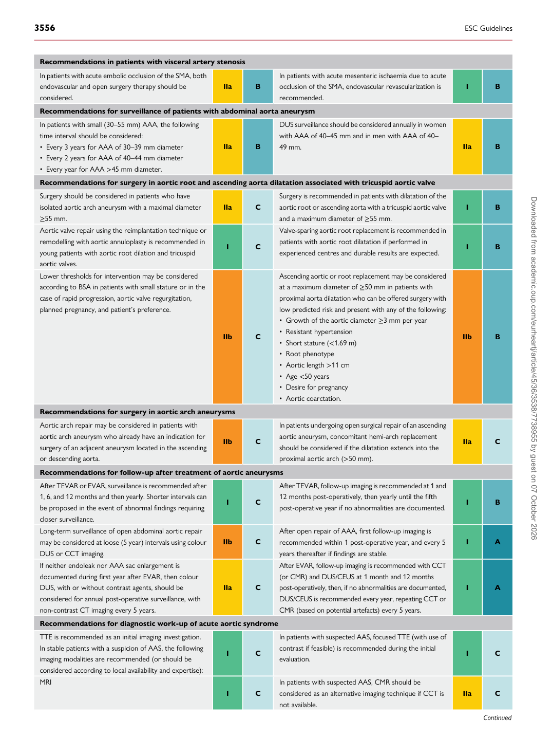
Recommendations in patients with visceral artery stenosis In patients with acute embolic occlusion of the SMA, both endovascular and open surgery therapy should be considered. IIa B In patients with acute mesenteric ischaemia due to acute occlusion of the SMA, endovascular revascularization is

recommended. I B

Recommendations for surveillance of patients with abdominal aorta aneurysm In patients with small (30–55 mm) AAA, the following time interval should be considered: • Every 3 years for AAA of 30–39 mm diameter • Every 2 years for AAA of 40–44 mm diameter • Every year for AAA >45 mm diameter. IIa B DUS surveillance should be considered annually in women with AAA of 40–45 mm and in men with AAA of 40–

49 mm. IIa B

Recommendations for surgery in aortic root and ascending aorta dilatation associated with tricuspid aortic valve Surgery should be considered in patients who have isolated aortic arch aneurysm with a maximal diameter ≥55 mm. IIa C Surgery is recommended in patients with dilatation of the aortic root or ascending aorta with a tricuspid aortic valve

and a maximum diameter of ≥55 mm. I B

Aortic valve repair using the reimplantation technique or remodelling with aortic annuloplasty is recommended in young patients with aortic root dilation and tricuspid aortic valves. I C Valve-sparing aortic root replacement is recommended in

patients with aortic root dilatation if performed in experienced centres and durable results are expected. I B

Lower thresholds for intervention may be considered according to BSA in patients with small stature or in the case of rapid progression, aortic valve regurgitation, planned pregnancy, and patient’s preference.

IIb C Ascending aortic or root replacement may be considered at a maximum diameter of ≥50 mm in patients with

proximal aorta dilatation who can be offered surgery with low predicted risk and present with any of the following: • Growth of the aortic diameter ≥3 mm per year • Resistant hypertension • Short stature (<1.69 m) • Root phenotype • Aortic length >11 cm • Age <50 years • Desire for pregnancy • Aortic coarctation. IIb B

Recommendations for surgery in aortic arch aneurysms Aortic arch repair may be considered in patients with aortic arch aneurysm who already have an indication for surgery of an adjacent aneurysm located in the ascending or descending aorta. IIb C In patients undergoing open surgical repair of an ascending

aortic aneurysm, concomitant hemi-arch replacement should be considered if the dilatation extends into the proximal aortic arch (>50 mm). IIa C

Recommendations for follow-up after treatment of aortic aneurysms After TEVAR or EVAR, surveillance is recommended after 1, 6, and 12 months and then yearly. Shorter intervals can be proposed in the event of abnormal findings requiring closer surveillance. I C After TEVAR, follow-up imaging is recommended at 1 and

12 months post-operatively, then yearly until the fifth post-operative year if no abnormalities are documented. I B

Long-term surveillance of open abdominal aortic repair may be considered at loose (5 year) intervals using colour DUS or CCT imaging. IIb C After open repair of AAA, first follow-up imaging is

recommended within 1 post-operative year, and every 5 years thereafter if findings are stable. I A

If neither endoleak nor AAA sac enlargement is documented during first year after EVAR, then colour DUS, with or without contrast agents, should be considered for annual post-operative surveillance, with non-contrast CT imaging every 5 years. IIa C After EVAR, follow-up imaging is recommended with CCT

(or CMR) and DUS/CEUS at 1 month and 12 months post-operatively, then, if no abnormalities are documented, DUS/CEUS is recommended every year, repeating CCT or CMR (based on potential artefacts) every 5 years. I A

Recommendations for diagnostic work-up of acute aortic syndrome TTE is recommended as an initial imaging investigation. In stable patients with a suspicion of AAS, the following imaging modalities are recommended (or should be considered according to local availability and expertise): I C In patients with suspected AAS, focused TTE (with use of

contrast if feasible) is recommended during the initial evaluation. I C

MRI

I C In patients with suspected AAS, CMR should be

considered as an alternative imaging technique if CCT is not available. IIa C

Downloaded from academic.oup.com/eurheartj/article/45/36/3538/7738955 by guest on 07 October 2026