p.124 抽出テキスト
16 Evidence tables

Evidence tables are available on the European Heart Journal website.

17 Data availability statement

No new data were generated or analysed in support of this research.

18 Author information

Author/task force Member Affiliations: Gisela Teixido-Tura, Cardiovascular Imaging section and Aortic diseases Unit (VASCERN), Department of Cardiology, Vall d’Hebron Hospital Universitari, Vall d’Hebron Barcelona Hospital Campus, Vall d′ Hebron Institut de Recerca (VHIR), Barcelona, Spain, Centro de Investigación Biomédica en Red de Enfermedades Cardiovasculares, Instituto de Salud Carlos III, Madrid, Spain; Stefano Lanzi, Department of Angiology, Lausanne University Hospital, University of Lausanne, Lausanne, Switzerland; Vinko Boc, Department of Vascular Diseases, University Medical Centre Ljubljana, Ljubljana, Slovenia; Eduardo Bossone, Department of Public Health, University of Naples “Federico II”, Naples, Italy, Department of Translational Medical Sciences, University of Naples “Federico II”, Naples, Italy; Marianne Brodmann, Division of Angiology, Medical

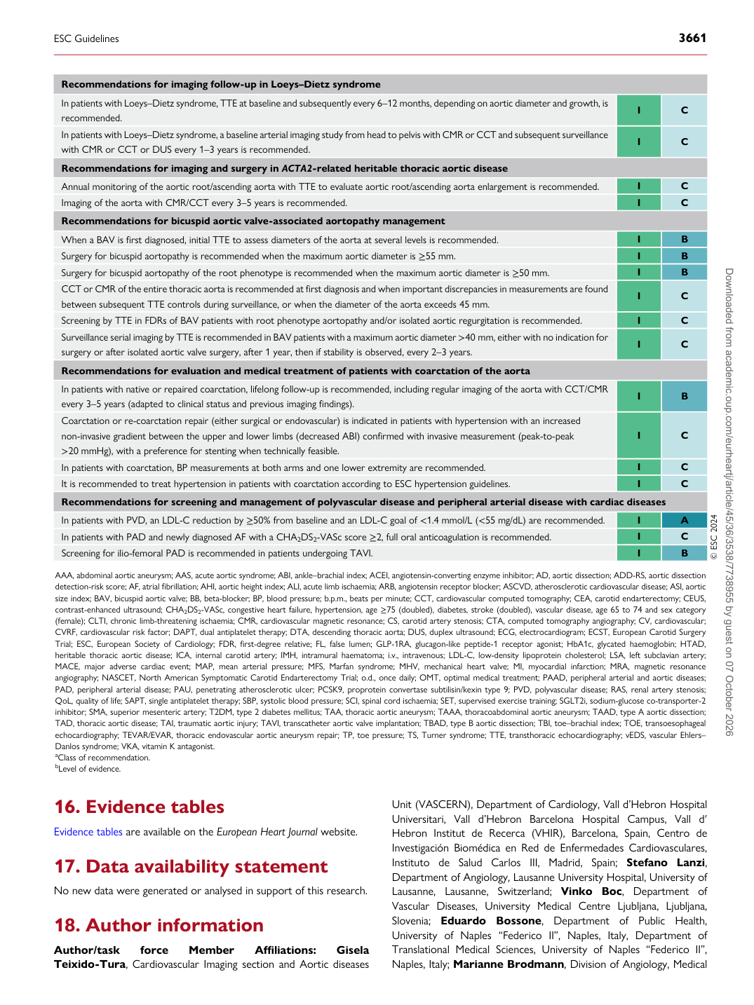
Recommendations for imaging follow-up in Loeys–Dietz syndrome In patients with Loeys–Dietz syndrome, TTE at baseline and subsequently every 6–12 months, depending on aortic diameter and growth, is recommended. I C In patients with Loeys–Dietz syndrome, a baseline arterial imaging study from head to pelvis with CMR or CCT and subsequent surveillance with CMR or CCT or DUS every 1–3 years is recommended. I C Recommendations for imaging and surgery in ACTA2-related heritable thoracic aortic disease Annual monitoring of the aortic root/ascending aorta with TTE to evaluate aortic root/ascending aorta enlargement is recommended. I C Imaging of the aorta with CMR/CCT every 3–5 years is recommended. I C Recommendations for bicuspid aortic valve-associated aortopathy management When a BAV is first diagnosed, initial TTE to assess diameters of the aorta at several levels is recommended. I B Surgery for bicuspid aortopathy is recommended when the maximum aortic diameter is ≥55 mm. I B Surgery for bicuspid aortopathy of the root phenotype is recommended when the maximum aortic diameter is ≥50 mm. I B CCT or CMR of the entire thoracic aorta is recommended at first diagnosis and when important discrepancies in measurements are found between subsequent TTE controls during surveillance, or when the diameter of the aorta exceeds 45 mm. I C Screening by TTE in FDRs of BAV patients with root phenotype aortopathy and/or isolated aortic regurgitation is recommended. I C Surveillance serial imaging by TTE is recommended in BAV patients with a maximum aortic diameter >40 mm, either with no indication for surgery or after isolated aortic valve surgery, after 1 year, then if stability is observed, every 2–3 years. I C Recommendations for evaluation and medical treatment of patients with coarctation of the aorta In patients with native or repaired coarctation, lifelong follow-up is recommended, including regular imaging of the aorta with CCT/CMR every 3–5 years (adapted to clinical status and previous imaging findings). I B Coarctation or re-coarctation repair (either surgical or endovascular) is indicated in patients with hypertension with an increased non-invasive gradient between the upper and lower limbs (decreased ABI) confirmed with invasive measurement (peak-to-peak >20 mmHg), with a preference for stenting when technically feasible. I C In patients with coarctation, BP measurements at both arms and one lower extremity are recommended. I C It is recommended to treat hypertension in patients with coarctation according to ESC hypertension guidelines. I C Recommendations for screening and management of polyvascular disease and peripheral arterial disease with cardiac diseases In patients with PVD, an LDL-C reduction by ≥50% from baseline and an LDL-C goal of <1.4 mmol/L (<55 mg/dL) are recommended. I A In patients with PAD and newly diagnosed AF with a CHA2DS2-VASc score ≥2, full oral anticoagulation is recommended. I C Screening for ilio-femoral PAD is recommended in patients undergoing TAVI. I B © ESC 2024

AAA, abdominal aortic aneurysm; AAS, acute aortic syndrome; ABI, ankle–brachial index; ACEI, angiotensin-converting enzyme inhibitor; AD, aortic dissection; ADD-RS, aortic dissection detection-risk score; AF, atrial fibrillation; AHI, aortic height index; ALI, acute limb ischaemia; ARB, angiotensin receptor blocker; ASCVD, atherosclerotic cardiovascular disease; ASI, aortic size index; BAV, bicuspid aortic valve; BB, beta-blocker; BP, blood pressure; b.p.m., beats per minute; CCT, cardiovascular computed tomography; CEA, carotid endarterectomy; CEUS, contrast-enhanced ultrasound; CHA2DS2-VASc, congestive heart failure, hypertension, age ≥75 (doubled), diabetes, stroke (doubled), vascular disease, age 65 to 74 and sex category (female); CLTI, chronic limb-threatening ischaemia; CMR, cardiovascular magnetic resonance; CS, carotid artery stenosis; CTA, computed tomography angiography; CV, cardiovascular; CVRF, cardiovascular risk factor; DAPT, dual antiplatelet therapy; DTA, descending thoracic aorta; DUS, duplex ultrasound; ECG, electrocardiogram; ECST, European Carotid Surgery Trial; ESC, European Society of Cardiology; FDR, first-degree relative; FL, false lumen; GLP-1RA, glucagon-like peptide-1 receptor agonist; HbA1c, glycated haemoglobin; HTAD, heritable thoracic aortic disease; ICA, internal carotid artery; IMH, intramural haematoma; i.v., intravenous; LDL-C, low-density lipoprotein cholesterol; LSA, left subclavian artery; MACE, major adverse cardiac event; MAP, mean arterial pressure; MFS, Marfan syndrome; MHV, mechanical heart valve; MI, myocardial infarction; MRA, magnetic resonance angiography; NASCET, North American Symptomatic Carotid Endarterectomy Trial; o.d., once daily; OMT, optimal medical treatment; PAAD, peripheral arterial and aortic diseases; PAD, peripheral arterial disease; PAU, penetrating atherosclerotic ulcer; PCSK9, proprotein convertase subtilisin/kexin type 9; PVD, polyvascular disease; RAS, renal artery stenosis; QoL, quality of life; SAPT, single antiplatelet therapy; SBP, systolic blood pressure; SCI, spinal cord ischaemia; SET, supervised exercise training; SGLT2i, sodium-glucose co-transporter-2 inhibitor; SMA, superior mesenteric artery; T2DM, type 2 diabetes mellitus; TAA, thoracic aortic aneurysm; TAAA, thoracoabdominal aortic aneurysm; TAAD, type A aortic dissection; TAD, thoracic aortic disease; TAI, traumatic aortic injury; TAVI, transcatheter aortic valve implantation; TBAD, type B aortic dissection; TBI, toe–brachial index; TOE, transoesophageal echocardiography; TEVAR/EVAR, thoracic endovascular aortic aneurysm repair; TP, toe pressure; TS, Turner syndrome; TTE, transthoracic echocardiography; vEDS, vascular Ehlers– Danlos syndrome; VKA, vitamin K antagonist. aClass of recommendation. bLevel of evidence.

ESC Guidelines                                                                                                                                                                                          3661

Downloaded from academic.oup.com/eurheartj/article/45/36/3538/7738955 by guest on 07 October 2026