p.113 抽出テキスト
10.3.2 Aortic arch anatomic variants

A type I arch, where the three great vessels directly arise from the aorta, is the most common form, occurring in about 70% of the population. The type II (bovine) arch is the most frequent variant: type II-A (9% of the population) has the left common carotid artery arising from the innominate artery, and type II-B (13% of the population) has both the innominate and left common carotid arteries originating from a common point on the aortic arch.1538,1539 Limited data suggest that a bovine arch is associated with a higher risk of aortic dilation and aortic events/complications.1540,1541 These variations are important to report as they can impact specific medical procedures and diagnostic interpretations.

10.3.3 Aberrant subclavian artery and Kommerell’s

diverticulum The most common variant is the aberrant right subclavian artery, where the right subclavian artery arises as the last branch of the aortic arch, usually after the left subclavian artery, and often passes behind the oesophagus through the mediastinum, potentially causing dysphagia lusoria, respiratory symptoms, or recurrent laryngeal nerve palsy. The less common variant, the aberrant left subclavian artery, is typically associated with congenital heart defects, such as a right aortic arch. However, in adulthood, both variations are often incidental findings.1542 Kommerell’s diverticulum is a remnant of the fourth dorsal aortic arch due to incomplete regression, found in 20%–60% of those with an aberrant subclavian artery.1543 Surgical intervention is advised for a diverticulum orifice >30 mm or combined diverticulum and adjacent descending aorta diameter >50 mm, or both.1544 Successful repair has been described using open, endovascular, or hybrid approaches depending on anatomy, comorbidities, and expertise.1543 11 Polyvascular peripheral arterial

disease and peripheral arterial disease in patients with cardiac diseases

11.1 Polyvascular disease

Polyvascular disease is defined as the simultaneous presence of clinically relevant obstructive atherosclerotic lesions in at least two major arterial territories.

11.1.1 Epidemiology and prognosis

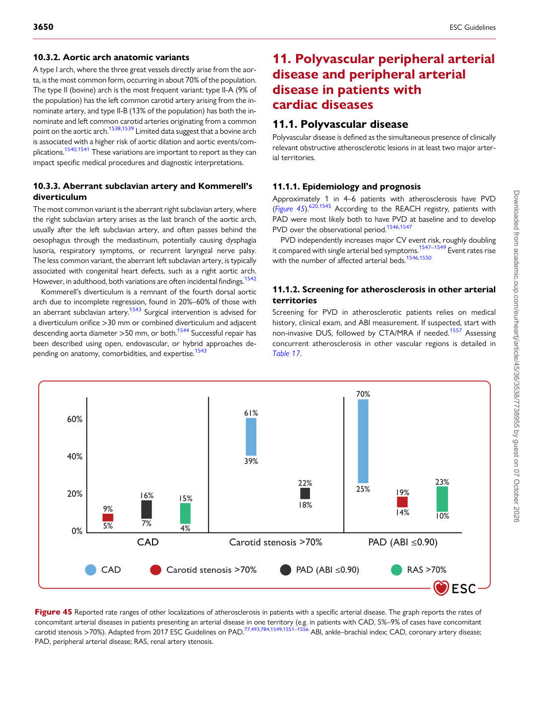
Approximately 1 in 4–6 patients with atherosclerosis have PVD (Figure 45).620,1545 According to the REACH registry, patients with PAD were most likely both to have PVD at baseline and to develop PVD over the observational period.1546,1547 PVD independently increases major CV event risk, roughly doubling it compared with single arterial bed symptoms.1547–1549 Event rates rise with the number of affected arterial beds.1546,1550

11.1.2 Screening for atherosclerosis in other arterial

territories Screening for PVD in atherosclerotic patients relies on medical history, clinical exam, and ABI measurement. If suspected, start with non-invasive DUS, followed by CTA/MRA if needed.1557 Assessing concurrent atherosclerosis in other vascular regions is detailed in Table 17.

CAD Carotid stenosis >70% PAD (ABI ≤0.90) RAS >70%

9% 5% 16% 4% 61%

39%

22%

18%

7% 15% 25%

70%

60%

40%

20%

0% 19%

14% 23%

10%

CAD Carotid stenosis >70% PAD (ABI ≤0.90)

Figure 45 Reported rate ranges of other localizations of atherosclerosis in patients with a specific arterial disease. The graph reports the rates of concomitant arterial diseases in patients presenting an arterial disease in one territory (e.g. in patients with CAD, 5%–9% of cases have concomitant carotid stenosis >70%). Adapted from 2017 ESC Guidelines on PAD.77,493,784,1549,1551–1556 ABI, ankle–brachial index; CAD, coronary artery disease; PAD, peripheral arterial disease; RAS, renal artery stenosis. Downloaded from academic.oup.com/eurheartj/article/45/36/3538/7738955 by guest on 07 October 2026