p.112 抽出テキスト
Y N

≥20 mmHg <20 mmHg

Perform catheterization (Re)coarctation

not likely

Surveillance: TTE every year and CMR or CCT 3–5 years

BP measurement and investigationa

Intervention

(Class IIa)

Intervention

(Class IIa)

Intervention

(Class I) BP measurement and investigationa

≥50% narrowing on any imaging modality

HTN HTN

N Y N

Y

N

Y

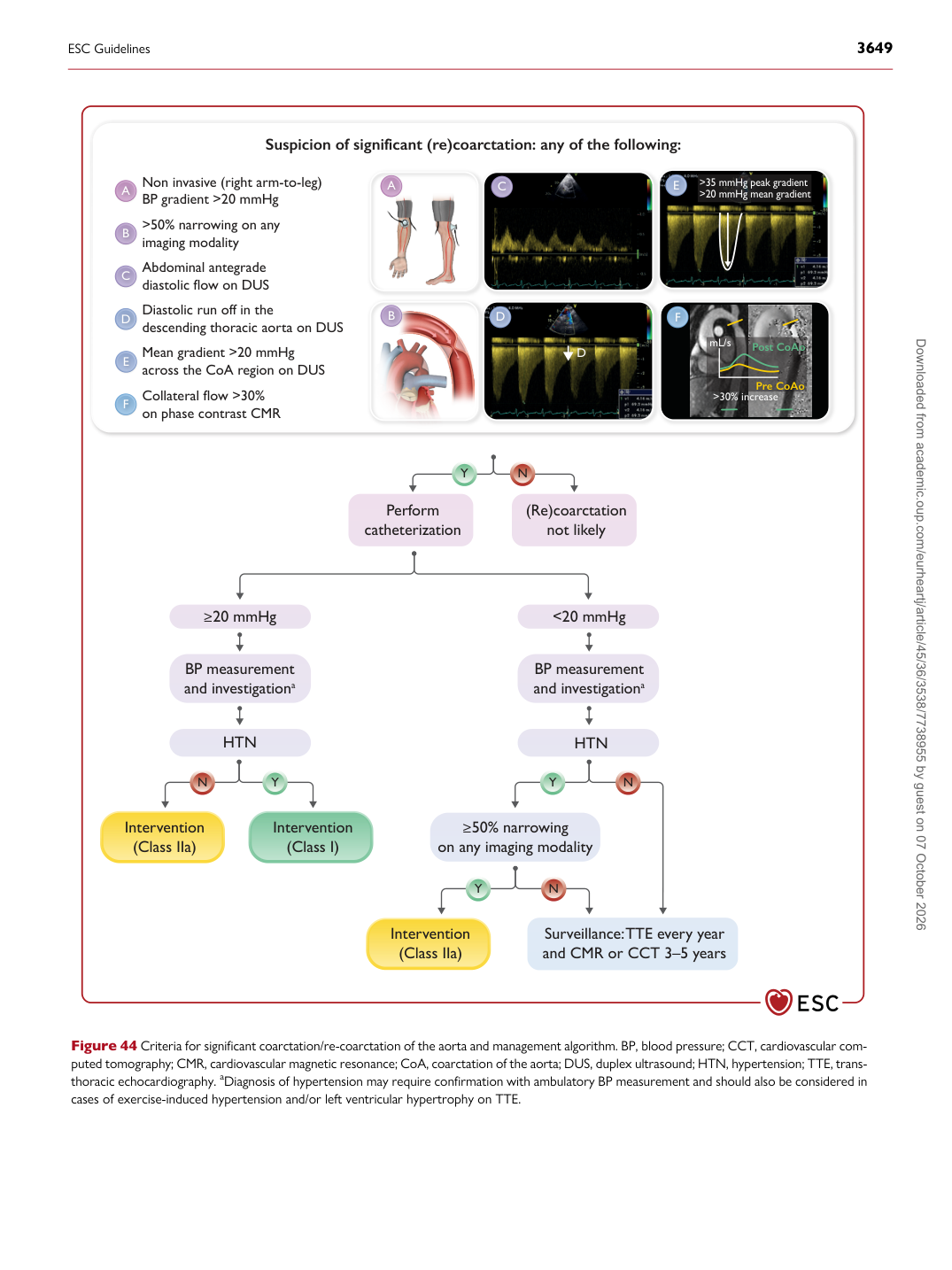
Suspicion of significant (re)coarctation: any of the following:

Non invasive (right arm-to-leg) BP gradient >20 mmHg >50% narrowing on any imaging modality Abdominal antegrade diastolic flow on DUS Diastolic run off in the descending thoracic aorta on DUS Mean gradient >20 mmHg across the CoA region on DUS Collateral flow >30% on phase contrast CMR

A A

B

B

C C

E E

D D

F F

D >35 mmHg peak gradient >20 mmHg mean gradient

Post CoAo

Pre CoAo

mL/s

>30% increase

Figure 44 Criteria for significant coarctation/re-coarctation of the aorta and management algorithm. BP, blood pressure; CCT, cardiovascular computed tomography; CMR, cardiovascular magnetic resonance; CoA, coarctation of the aorta; DUS, duplex ultrasound; HTN, hypertension; TTE, transthoracic echocardiography. aDiagnosis of hypertension may require confirmation with ambulatory BP measurement and should also be considered in cases of exercise-induced hypertension and/or left ventricular hypertrophy on TTE.

ESC Guidelines                                                                                                                                                                                          3649

Downloaded from academic.oup.com/eurheartj/article/45/36/3538/7738955 by guest on 07 October 2026