p.111 抽出テキスト
10.3 Coarctation of the aorta and aortic

arch variants 10.3.1. Coarctation of the aorta This topic is extensively discussed in the ESC 2020 Guidelines for the management of adult congenital heart disease.1468 Coarctation of the aorta (CoA) manifests as a discrete stenosis or a hypoplastic segment typically located at the insertion of the ductus arteriosus. More distal locations are known as mid-aortic syndrome and require dedicated management.1524 Associated lesions include BAV (up to 50%–85%), intracerebral aneurysms (10%), and ascending aortic aneurysms.1525,1526 CoA may be associated with syndromes such as TS. Research indicates that up to 12.6% of females diagnosed with CoA also have TS, and coarctation is observed in 7%–18% of patients with TS.1417,1468,1527

10.3.1.1 Diagnostic work-up

Mild cases of CoA may only become evident in adulthood. Symptoms reflect pre-stenotic hypertension (e.g. headache, nosebleeds) and poststenotic hypoperfusion (e.g. abdominal angina and claudication). The natural course is largely driven by hypertension-related complications, including HF, intracranial haemorrhage, premature coronary/cerebral artery disease, and aortic rupture/dissection.1528 Presently, there is no evidence supporting screening for intracerebral aneurysms in asymptomatic patients. A systolic non-invasive gradient between upper and lower extremities, an abnormal ABI, or an invasive peak-to-peak gradient ≥20 mmHg indicates significant CoA. In the presence of collaterals or decreased LV function, gradients or ABI may underestimate severity. A diastolic tail in the DTA or abdominal diastolic antegrade flow by TTE is suggestive of significant narrowing. Criteria to consider significant CoA are listed in Figure 44. TTE is also useful to detect LV hypertrophy, which is a marker of disease. CMR and CCT are the preferred imaging techniques, depicting the narrowing as well as the surrounding anatomy, necessary for interventional decision-making.

10.3.1.2 Treatment and follow-up

In native CoA and re-coarctation (Figure 44) covered stenting is the firstchoice treatment. Interposition of a tube graft is the preferred surgical therapy if stenting is less suitable.1529 Hypertension remains an important complication, even after successful treatment, and is more common when the initial repair is performed in adulthood.1528 Right arm 24 h ambulatory BP measurement or exercise tests better detect hypertension.1530,1531 All CoA patients require lifelong follow-up.1532 Imaging of the aorta with CMR/CCT every 3–5 years, adjusted to previous imaging findings and type of intervention, is required to document post-repair or post-interventional complications (such as re-coarctation). Patch repairs are at particular risk of repair-site para-anastomotic aneurysms or pseudo-aneurysms, the latter possibly occurring following interposition grafts as well.1533

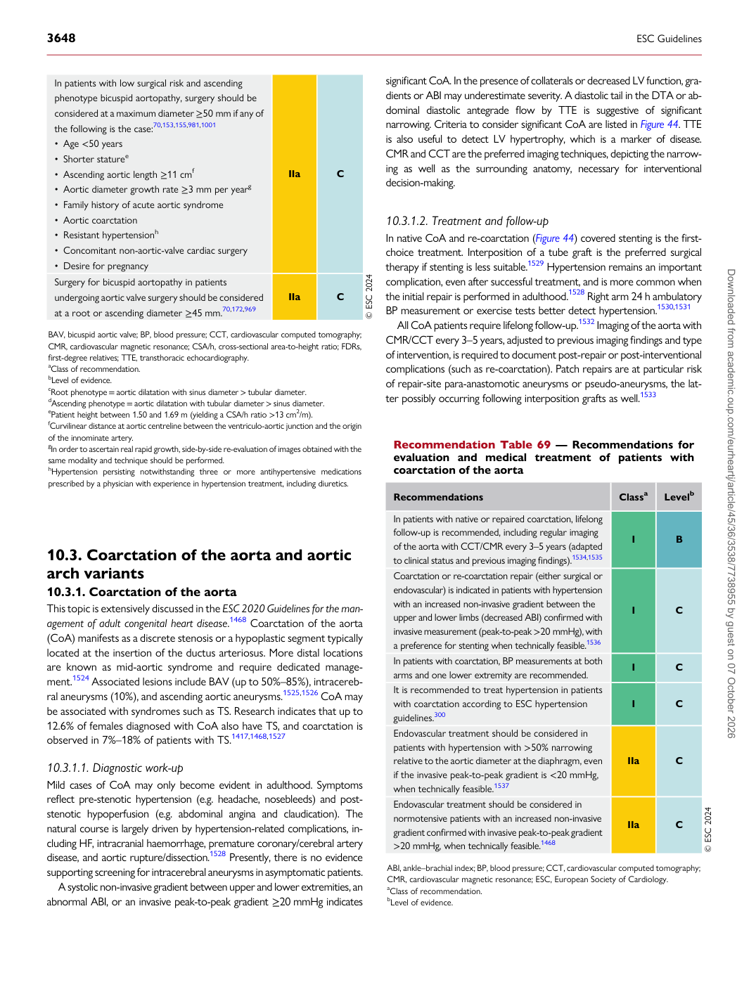
Recommendation Table 69 — Recommendations for evaluation and medical treatment of patients with coarctation of the aorta

Recommendations Classa Levelb

In patients with native or repaired coarctation, lifelong follow-up is recommended, including regular imaging of the aorta with CCT/CMR every 3–5 years (adapted to clinical status and previous imaging findings).1534,1535 I B Coarctation or re-coarctation repair (either surgical or endovascular) is indicated in patients with hypertension with an increased non-invasive gradient between the upper and lower limbs (decreased ABI) confirmed with invasive measurement (peak-to-peak >20 mmHg), with a preference for stenting when technically feasible.1536 I C In patients with coarctation, BP measurements at both arms and one lower extremity are recommended. I C It is recommended to treat hypertension in patients with coarctation according to ESC hypertension guidelines.300 I C Endovascular treatment should be considered in patients with hypertension with >50% narrowing relative to the aortic diameter at the diaphragm, even if the invasive peak-to-peak gradient is <20 mmHg, when technically feasible.1537 IIa C Endovascular treatment should be considered in normotensive patients with an increased non-invasive gradient confirmed with invasive peak-to-peak gradient >20 mmHg, when technically feasible.1468 IIa C © ESC 2024

ABI, ankle–brachial index; BP, blood pressure; CCT, cardiovascular computed tomography; CMR, cardiovascular magnetic resonance; ESC, European Society of Cardiology. aClass of recommendation. bLevel of evidence.

In patients with low surgical risk and ascending phenotype bicuspid aortopathy, surgery should be considered at a maximum diameter ≥50 mm if any of the following is the case:70,153,155,981,1001 • Age <50 years • Shorter staturee • Ascending aortic length ≥11 cmf • Aortic diameter growth rate ≥3 mm per yearg • Family history of acute aortic syndrome • Aortic coarctation • Resistant hypertensionh • Concomitant non-aortic-valve cardiac surgery • Desire for pregnancy IIa C Surgery for bicuspid aortopathy in patients undergoing aortic valve surgery should be considered at a root or ascending diameter ≥45 mm.70,172,969 IIa C © ESC 2024

BAV, bicuspid aortic valve; BP, blood pressure; CCT, cardiovascular computed tomography; CMR, cardiovascular magnetic resonance; CSA/h, cross-sectional area-to-height ratio; FDRs, first-degree relatives; TTE, transthoracic echocardiography. aClass of recommendation. bLevel of evidence. cRoot phenotype = aortic dilatation with sinus diameter > tubular diameter. dAscending phenotype = aortic dilatation with tubular diameter > sinus diameter. ePatient height between 1.50 and 1.69 m (yielding a CSA/h ratio >13 cm2/m). fCurvilinear distance at aortic centreline between the ventriculo-aortic junction and the origin of the innominate artery. gIn order to ascertain real rapid growth, side-by-side re-evaluation of images obtained with the same modality and technique should be performed. hHypertension persisting notwithstanding three or more antihypertensive medications prescribed by a physician with experience in hypertension treatment, including diuretics. Downloaded from academic.oup.com/eurheartj/article/45/36/3538/7738955 by guest on 07 October 2026