p.104 抽出テキスト
10.1.2 Vascular Ehlers–Danlos syndrome

10.1.2.1. Diagnosis, clinical presentation, and natural history Vascular Ehlers–Danlos syndrome is a rare (prevalence of 1/50 000 to 1/200 000) autosomal dominant disease caused by pathogenic variants in the COL3A1 gene, which encodes the pro-alpha1 chains of type III procollagen. The most common COL3A1 variants provoke a disruption in the assembly of type III collagen fibrils, causing an important loss of mechanical strength of arteries and other hollow organs, especially the bowel and uterus.1438 Identification of a causal COL3A1 variant is a requirement for the diagnosis of vEDS.1439 vEDS is the most severe form of Ehlers–Danlos syndrome because of its clinical life-threatening vascular complications, making early identification and a thorough family inquiry particularly crucial. Clinical complications may start during adolescence and repeat at unpredictable time intervals. The most common complications involve medium-sized arteries: dissections, aneurysms, arterial ruptures, and arteriovenous fistulas. AD (both type A and B) occurs in up to 10% of patients.1440 Prognosis depends on the type of COL3A1 variant, with null variants (no gene product or absence of function) showing a better outcome.1441 The rate of recurrence of organic complications in patients with vEDS is 1.6 events per 5 year period. Life expectancy is reduced to an average of 51 years.1442

10.1.2.2 Surveillance and imaging

Management of vEDS is complex and requires a multidisciplinary approach. Recommendations include: lifestyle modification to minimize injury and risk of vessel/organ rupture, identification of a care team, individualized emergency care plans, maintaining BP in the normal range, aggressive hypertension treatment, and annual surveillance of the vascular tree by DUS, CCT (low radiation alternatives), or CMR (if feasible).1439 A recent survey among European expert centres indicated that arterial monitoring is standard clinical practice and that frequency of follow-up should be adapted individually.1443 The prognosis improves when patients are properly managed.1441

10.1.2.3 Medical treatment

Medical management is based on optimal BP control. Celiprolol, a BB with vasodilatory properties, has been shown to reduce vascular morbidity in two retrospective studies1441,1444 and one randomized, openlabel trial.1445 There is no consensus about the age at which to start treatment, but starting after 10 years of age is considered reasonable by many experts. 10.1.2.4 Surgical treatment

Acute, unexplained pain requires urgent imaging to exclude arterial rupture. Acute arterial complications usually require hospitalization and a conservative approach in most cases. Interventional vascular or intestinal procedures are limited to vital risk. Procedures requiring organ inflation should be avoided or performed with extreme caution. There are no clear recommendations regarding aortic/arterial diameters at which to intervene in patients with vEDS. Thus, decisions need to be made on a case-by-case basis.

10.1.2.5 Pregnancy

Pregnancy in vEDS incurs a risk of (fatal) arterial and uterine complications. Pregnancy does not appear to affect overall mortality compared with nulliparous vEDS women.1446 However, patients need to be engaged in a shared-decision process, informed by vascular status and underlying variant type.

10.1.3 Marfan syndrome

10.1.3.1. Diagnosis, clinical presentation, and natural history Marfan syndrome, the most common syndromic HTAD condition (prevalence of 1/5000–1/10 000), arises from pathogenic fibrillin-1 gene (FBN1) variants. Beyond the CV system, multiple organ systems are often affected, including the eyes and skeleton. Diagnosis relies on recognizing clinical features in line with the revised Ghent nosology, which includes genetic testing.1447 Aortic aneurysm and dissection involving the aortic root are a hallmark of the disease. Less commonly, the descending thoracic and abdominal aorta may be involved. With increasing survival and age in MFS, the prevalence of TBAD seems to be increasing, exceeding type A dissection rates in recent reports.1448,1449 TBAD will often occur at diameters below surgical thresholds. Previous aortic root replacement, mitral valve surgery, and a longer life span are associated with TBAD. Additional CV features include mitral valve prolapse, extra-aortic arterial involvement, myocardial dysfunction, and arrhythmias.1393,1450–1452 Thanks to improved diagnosis in earlier stages, proper management including surveillance, medical treatment, and timely prophylactic aortic surgery, life expectancy in MFS patients is now approaching that of the general population.1416,1453 The major determinant of TAAD is the aortic root diameter, with increased risk of rupture when it exceeds 50 mm.1454 Other risk factors include family history of AAS at low diameter, aortic root growth rate (annualized growth rate ≥3 mm or more in adults), pregnancy, and hypertension (hypertension persisting notwithstanding three or more antihypertensive medications prescribed by a physician with experience in hypertension treatment). Increasing evidence for variant-based differences in aortic risk is emerging and may be considered.1413,1416

10.1.3.2 Imaging surveillance

Transthoracic echocardiography is the appropriate imaging modality for initial evaluation and follow-up of the aortic root in most patients and allows evaluation of the distal segments of the aorta in many. Also, TTE is useful for assessing mitral and aortic valve regurgitation, mitral valve prolapse with/out annular disjunction, and LV dysfunction. In some cases (especially when pectus abnormalities are present) TTE windows may be suboptimal, and CMR (preferably)/CCT may be preferred. Periodical evaluation of the global aorta and peripheral arteries with CMR/CCT and DUS (every 3–5 years based on the patient’s evolution) is indicated since they also present a higher incidence of

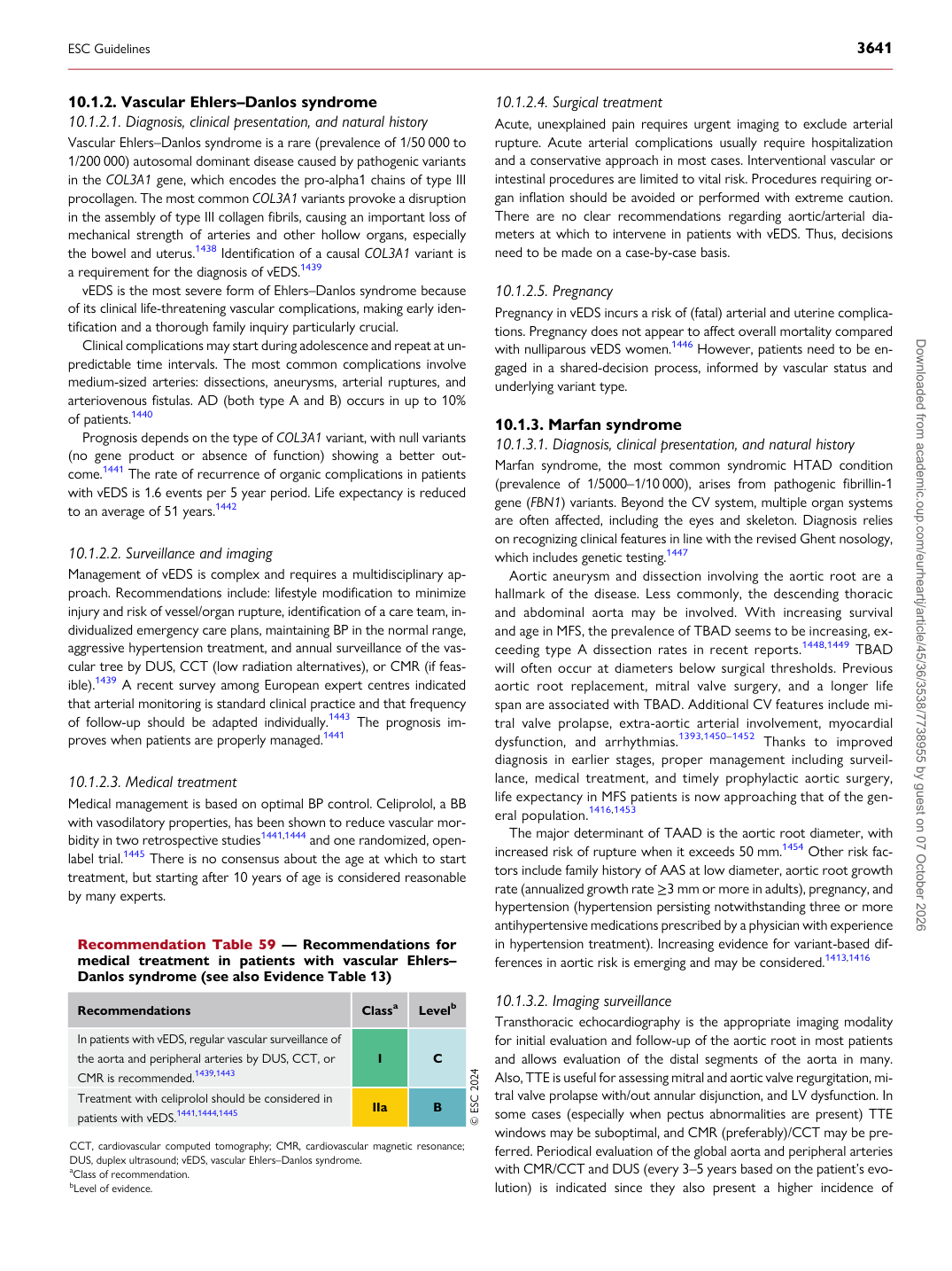
Recommendation Table 59 — Recommendations for medical treatment in patients with vascular Ehlers– Danlos syndrome (see also Evidence Table 13)

Recommendations Classa Levelb

In patients with vEDS, regular vascular surveillance of the aorta and peripheral arteries by DUS, CCT, or CMR is recommended.1439,1443 I C Treatment with celiprolol should be considered in patients with vEDS.1441,1444,1445 IIa B © ESC 2024

CCT, cardiovascular computed tomography; CMR, cardiovascular magnetic resonance; DUS, duplex ultrasound; vEDS, vascular Ehlers–Danlos syndrome. aClass of recommendation. bLevel of evidence.

ESC Guidelines                                                                                                                                                                                          3641

Downloaded from academic.oup.com/eurheartj/article/45/36/3538/7738955 by guest on 07 October 2026