p.101 抽出テキスト
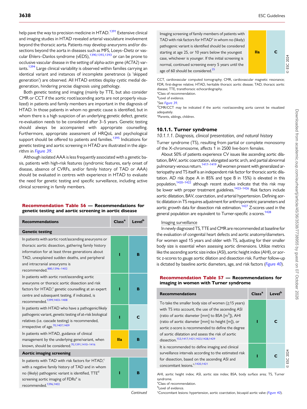
help pave the way to precision medicine in HTAD.1391 Extensive clinical and imaging studies in HTAD revealed arterial vasculature involvement beyond the thoracic aorta. Patients may develop aneurysms and/or dissections beyond the aorta in diseases such as MFS, Loeys–Dietz or vascular Ehlers–Danlos syndrome (vEDS),1390,1392,1393 or can be prone to occlusive vascular disease in the setting of alpha-actin gene (ACTA2) variants.1394 Large clinical variability is observed within families carrying an identical variant and instances of incomplete penetrance (a ‘skipped generation’) are observed. All HTAD entities display cystic medial degeneration, hindering precise diagnosis using pathology. Both genetic testing and imaging (mainly by TTE, but also consider CMR or CCT if the aortic root/ascending aorta are not properly visualized) in patients and family members are important in the diagnosis of HTAD. In those patients in whom no genetic cause is identified, but in whom there is a high suspicion of an underlying genetic defect, genetic re-evaluation needs to be considered after 3–5 years. Genetic testing should always be accompanied with appropriate counselling. Furthermore, appropriate assessment of HRQoL and psychological support should be offered to patients and families.1395 Indications for genetic testing and aortic screening in HTAD are illustrated in the algorithm in Figure 39. Although isolated AAA is less frequently associated with a genetic basis, patients with high-risk features (syndromic features, early onset of disease, absence of CVRFs, and/or family history of TAD or AAA) should be evaluated in centres with experience in HTAD to evaluate the need for genetic testing and specific surveillance, including active clinical screening in family members. 10.1.1 Turner syndrome

10.1.1.1. Diagnosis, clinical presentation, and natural history Turner syndrome (TS), resulting from partial or complete monosomy of the X-chromosome, affects 1 in 2500 live-born females. About 50% of patients experience CV issues like ascending aortic dilatation, BAV, aortic coarctation, elongated aortic arch, and partial abnormal pulmonary venous return.1417–1419 All women present with generalized arteriopathy and TS itself is an independent risk factor for thoracic aortic dilatation. AD risk (type A in 85% and type B in 15%) is elevated in this population,1420–1422 although recent studies indicate that this risk may be lower with proper treatment guidelines.1423–1426 Risk factors include aortic dilatation, BAV, coarctation, and arterial hypertension. Defining aortic dilatation in TS requires adjustment for anthropometric parameters and aortic growth data for dissection risk estimation.1427 Z-scores used in the general population are equivalent to Turner-specific z-scores.1428 Imaging surveillance In newly diagnosed TS, TTE and CMR are recommended at baseline for the evaluation of congenital heart defects and aortic anatomy/diameters. For women aged 15 years and older with TS, adjusting for their smaller body size is essential when assessing aortic dimensions. Utilize metrics like the ascending aortic size index (ASI), aortic height index (AHI), or aortic z-scores to gauge aortic dilation and dissection risk. Further follow-up is dictated by baseline aortic diameters, age, and risk factors (Figure 40).

Recommendation Table 57 — Recommendations for imaging in women with Turner syndrome

Recommendations Classa Levelb

To take the smaller body size of women (≥15 years) with TS into account, the use of the ascending ASI (ratio of aortic diameter [mm] to BSA [m2]), AHI (ratio of aortic diameter [mm] to height [m]), or aortic z-score is recommended to define the degree of aortic dilatation and assess the risk of aortic dissection.153,1417,1421,1423,1428,1429 I C It is recommended to define imaging and clinical surveillance intervals according to the estimated risk for dissection, based on the ascending ASI and concomitant lesions.c,1420,1421 I C © ESC 2024

AHI, aortic height index; ASI, aortic size index; BSA, body surface area; TS, Turner syndrome. aClass of recommendation. bLevel of evidence. cConcomitant lesions: hypertension, aortic coarctation, bicuspid aortic valve (Figure 40).

Recommendation Table 56 — Recommendations for genetic testing and aortic screening in aortic disease

Recommendations Classa Levelb

Genetic testing In patients with aortic root/ascending aneurysms or thoracic aortic dissection, gathering family history information for at least three generations about TAD, unexplained sudden deaths, and peripheral and intracranial aneurysms is recommended.880,1396–1402 I B In patients with aortic root/ascending aortic aneurysms or thoracic aortic dissection and risk factors for HTAD,c genetic counselling at an expert centre and subsequent testing, if indicated, is recommended.1399,1403–1408 I B In patients with HTAD who have a pathogenic/likely pathogenic variant, genetic testing of at-risk biological relatives (i.e. cascade testing) is recommended, irrespective of age.70,1407,1409 I C In patients with HTAD, guidance of clinical management by the underlying gene/variant, when known, should be considered.70,1391,1410–1416 IIa B Aortic imaging screening In patients with TAD with risk factors for HTAD,c with a negative family history of TAD and in whom no (likely) pathogenic variant is identified, TTEd screening aortic imaging of FDRse is recommended.1396,1402 I B

Imaging screening of family members of patients with TAD with risk factors for HTADc in whom no (likely) pathogenic variant is identified should be considered starting at age 25, or 10 years below the youngest case, whichever is younger. If the initial screening is normal, continued screening every 5 years until the age of 60 should be considered.25 IIa C © ESC 2024

CCT, cardiovascular computed tomography; CMR, cardiovascular magnetic resonance; FDR, first-degree relative; HTAD, heritable thoracic aortic disease; TAD, thoracic aortic disease; TTE, transthoracic echocardiography. aClass of recommendation. bLevel of evidence. cSee Figure 39. dCMR/CCT may be indicated if the aortic root/ascending aorta cannot be visualized adequately. eParents, siblings, children. Downloaded from academic.oup.com/eurheartj/article/45/36/3538/7738955 by guest on 07 October 2026