p.100 抽出テキスト
Type B IMH and PAU are usually conservatively treated with antihypertensive therapy and watchful monitoring. Most of the medically treated IMHs have a favourable course, whereas PAUs are less predictable in terms of risk of acute TBAD or rupture.1350 Therefore, for IMH the same surveillance criteria as for medically treated uncomplicated TBAD can be employed; for PAU more frequent controls are advisable, i.e. one every 6 months instead of every year. Selectively, in asymptomatic patients with 2 year growth-rate stabilization and no high-risk features, intervals between controls can be longer (every 1–2 years) (Figures 35 and 38).70,1384 10 Genetic and congenital diseases

of the aorta

10.1 Genetic and chromosomal diseases

This section discusses genetic and congenital aortic diseases. Aortic root and ascending aortic disease is commonly linked to congenital or hereditary factors, while descending aortic problems, especially in the AA, often result from atherosclerosis.1385 Unless noted otherwise, recommendations provided herein are intended for adults. Genetic diseases affecting the thoracic aorta are grouped under the broader term of HTAD. HTAD comprises a clinically and genetically heterogeneous group of disorders sharing the common denominator of aneurysm or dissection of the thoracic aorta. Familial forms (thoracic aortic disease [TAD] affecting ≥2 individuals in one family) or confirmed genetic entities (familial or sporadic) as well as syndromes conferring a risk for TAD fall under the definition of HTAD.70 Due to the rarity of these conditions, robust evidence for many scenarios, such as intervention thresholds, surgical methods, open surgery vs. endovascular approaches, and pregnancy planning, is lacking. Thus, a multidisciplinary and individualized approach is advisable.70,1386,1387

Clinically, HTADs can manifest as either syndromic or nonsyndromic entities. The genes identified to date may underly both entities and predominantly show autosomal dominant inheritance patterns. While TAD is the primary feature in HTAD, extra-aortic features (skeletal/ocular) may be key to diagnosing certain syndromic cases. In some cases, the presence of extra-aortic manifestations may aid in risk stratification and hence in defining optimal management.1388–1390 The main clinical and genetic data on syndromic and non-syndromic HTADs are summarized in the Supplementary data online, Table S5. Numerous underlying gene defects have been discovered in both syndromic and non-syndromic cases, leading to the constitution of three major molecular groups: genes encoding components of: (i) the extracellular matrix; (ii) the transforming growth factor-beta (TGF-ß) signalling pathway; and (iii) the smooth muscle cell contractile apparatus. Clinical and CV outcomes vary between these groups and will

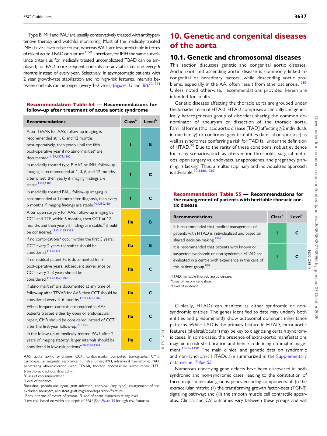
Recommendation Table 54 — Recommendations for follow-up after treatment of acute aortic syndrome

Recommendations Classa Levelb

After TEVAR for AAS, follow-up imaging is recommended at 1, 6, and 12 months post-operatively, then yearly until the fifth post-operative year if no abnormalitiesc are documented.1159,1378,1382 I B In medically treated type B AAS or IMH, follow-up imaging is recommended at 1, 3, 6, and 12 months after onset, then yearly if imaging findings are stable.1301,1383 I C In medically treated PAU, follow-up imaging is recommended at 1 month after diagnosis, then every

6 months if imaging findings are stable.70,1350,1384 I C After open surgery for AAS, follow-up imaging by CCT and TTE within 6 months, then CCT at 12 months and then yearly if findings are stable,d should be considered.1153,1159,1383 IIa B If no complicationsc occur within the first 5 years, CCT every 2 years thereafter should be considered.1159,1378 IIa B If no residual patent FL is documented for 3 post-operative years, subsequent surveillance by CCT every 2–3 years should be considered.1153,1159,1383 IIa C If abnormalitiesc are documented at any time of follow-up after TEVAR for AAS, then CCT should be considered every 3–6 months.1159,1378,1382 IIa C When frequent controls are required in AAS patients treated either by open or endovascular repair, CMR should be considered instead of CCT after the first-year follow-up.70,1153 IIa C In the follow-up of medically treated PAU, after 2 years of imaging stability, larger intervals should be considered in low-risk patientse.70,1350,1384 IIa C © ESC 2024

AAS, acute aortic syndrome; CCT, cardiovascular computed tomography; CMR, cardiovascular magnetic resonance; FL, false lumen; IMH, intramural haematoma; PAU, penetrating atherosclerotic ulcer; TEVAR, thoracic endovascular aortic repair; TTE, transthoracic echocardiography. aClass of recommendation. bLevel of evidence. cIncluding: pseudo-aneurysm, graft infection, endoleak (any type), enlargement of the excluded aneurysm, and stent graft migration/separation/fracture. dBoth in terms of extent of residual FL and of aortic diameters at any level. eLow-risk: based on width and depth of PAU (See Figure 35 for high-risk features). Recommendation Table 55 — Recommendations for the management of patients with heritable thoracic aortic disease

Recommendations Classa Levelb

It is recommended that medical management of patients with HTAD is individualized and based on shared decision-making.1386 I C It is recommended that patients with known or suspected syndromic or non-syndromic HTAD are evaluated in a centre with experience in the care of this patient group.888 I C © ESC 2024

HTAD, heritable thoracic aortic disease. aClass of recommendation. bLevel of evidence.

ESC Guidelines                                                                                                                                                                                          3637

Downloaded from academic.oup.com/eurheartj/article/45/36/3538/7738955 by guest on 07 October 2026