p.72 抽出テキスト
9.14.4 Phaeochromocytoma/paraganglioma

Phaeochromocytomas are rare adrenal tumours that secrete catecholamines and are present in <0.2% of patients with hypertension. A small percentage (<10%) of catecholamine-producing tumours are extraadrenal and are derived from sympathetic and non-sympathetic nerves. More than 35% of the non-syndromic PPGLs are due to germline mutations.338 These mutations should be screened for because, when found, they can drive management of the proband and the family and also inform the choice of functional imaging. Moreover, some germline mutations, such as those involving succinate dehydrogenase B, carry a risk of malignant adrenal tumours.301,336 Sympathetic PPGLs are usually secreting and present with chronic, episodic, or labile hypertension. Adrenergic crises cause hypertensive emergencies and should be treated with an intravenous (i.v.) alpha-1-blocker, such as phentolamine, doxazosin or terazosin, or labetalol. When given i.v. (1–2 mg/kg) twice weekly as a bolus over 1 min followed by a continuous infusion, labetalol also has alphablocker properties and has the advantage of allowing titration of the infusion based on the BP response, and avoids tachycardia via beta-blockade. Identifying a single tumour mandates surgical excision after adequate pharmacological preparation, because secreting PPGLs can cause fatal events with no warning. Administering doxazosin or terazosin, followed by a beta-blocker, usually controls BP and adrenergic crises. As PPGLs are associated with a redistribution of volume from the periphery to the cardiopulmonary system,944 patients with PPGLs have peripheral hypovolaemia that exposes them to the risk of profound hypotension, particularly right after tumour excision. Therefore, adequate fluid administration should be carefully managed.

9.14.5 Obstructive sleep apnoea syndrome

The management of this prevalent condition should be driven by the result of a polysomnography study, which should provide the value of the AHI (the average number of episodes per hour) and the sleep position in which apnoeic–hypopnoeic episodes occur. For mild OSAS (AHI < 15), weight loss and advice on sleep hygiene are usually sufficient. For moderate (AHI of 15–30) and severe (AHI > 30) OSAS, continuous positive airway pressure (CPAP) is indicated and usually improves BP control and helps to resolve resistant hypertension. If CPAP is not tolerated, the site of upper airway obstruction should be determined by an Ear, Nose, and Throat evaluation with drug-induced sleep endoscopy as a potential step to corrective surgery. 9.14.6 Drug-induced hypertension

Over-the-counter medications, prescribed drugs, and drug abuse (recreational substances and misuse of drugs) can cause hypertension (Supplementary data online, Table S4).

9.14.6.1 Anticancer drug-induced hypertension

Growing evidence indicates that, while contemporary anticancer and anti-angiogenic drugs improve cancer survival, they can also cause hypertension (Supplementary data online, Table S4). This is especially evident in patients treated with vascular endothelial growth factor inhibitors, in whom BP increases in 80%–90%.945 Tyrosine kinase inhibitors and proteasome inhibitors also increase BP, as do adjuvant therapies (corticosteroids, calcineurin inhibitors, non-steroidal anti-inflammatory drugs, and anti-androgen hormone therapy). Hypertension caused by anticancer drugs is often dose limiting and may be reversible after therapy interruption or discontinuation. Evidence-based clinical trials specifically addressing patients who develop hypertension due to cancer therapy are lacking. It is recommended that management of hypertension in these patients follows that for the general population.945,946 Managing these complex patients requires multidisciplinary healthcare involving oncologists, hypertension specialists, cardiologists, and nephrologists,945,946 as highlighted in the 2022 ESC Guidelines on cardio-oncology.946

9.14.7 Other forms of secondary hypertension

Other forms of secondary hypertension, such as genetic causes of hypertension (Liddle’s syndrome, glucocorticoid-remediable aldosteronism), excess liquorice, Cushing’s syndrome, thyroid disease, hyperparathyroidism, aortic coarctation, and acromegaly are rare. Affected patients should be referred to specialized centres for treatment.

10 Acute and short-term lowering

of blood pressure

10.1 Acute blood pressure management in

hypertensive emergencies 10.1.1. Definition and characteristics of hypertensive emergencies Hypertensive emergency is defined as BP of ≥180/110 mmHg (see Figure 10) associated with acute HMOD, often in the presence of symptoms. Hypertensive emergencies are potentially life-threatening and require immediate and careful intervention to reduce BP, often with i.v. therapy. Symptoms of hypertensive emergency depend on the organs affected but may include headache, visual disturbances, chest pain, shortness of breath, dizziness, and other neurological deficits. In patients with hypertensive encephalopathy, somnolence, lethargy, tonic–clonic seizures, and cortical blindness may precede a loss of consciousness; however, focal neurological lesions are rare and should raise the suspicion of stroke. As outlined in Section 7, we define HMOD among patients with chronically elevated BP or hypertension as the presence of specific cardiac, vascular, and renal alterations.31,159 However, in the setting of hypertensive emergency, more acute manifestations of organ damage are relevant for management.

Acute manifestations of organ damage include: • Patients with severe acute hypertension associated with other clinical conditions likely to require urgent reduction in BP, e.g. acute onset of aortic dissection, myocardial ischaemia, eclampsia, or heart failure.

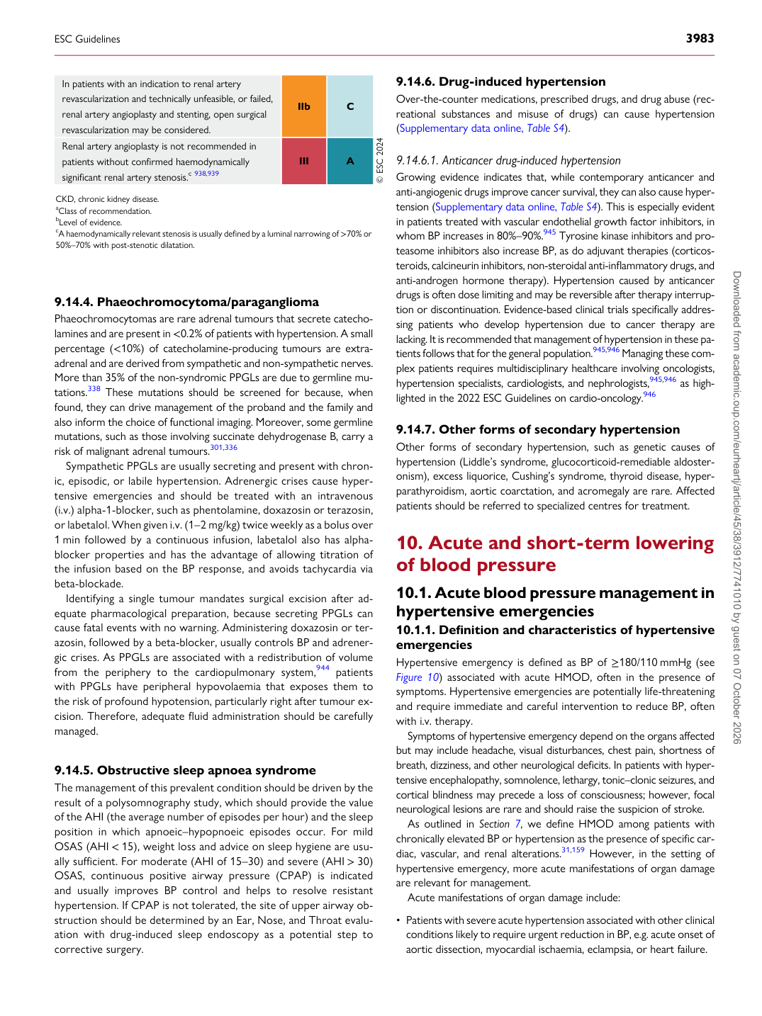
In patients with an indication to renal artery revascularization and technically unfeasible, or failed, renal artery angioplasty and stenting, open surgical revascularization may be considered. IIb C Renal artery angioplasty is not recommended in patients without confirmed haemodynamically significant renal artery stenosis.c 938,939 III A © ESC 2024

CKD, chronic kidney disease. aClass of recommendation. bLevel of evidence. cA haemodynamically relevant stenosis is usually defined by a luminal narrowing of >70% or 50%–70% with post-stenotic dilatation.

ESC Guidelines                                                                                                                                                                                          3983

Downloaded from academic.oup.com/eurheartj/article/45/38/3912/7741010 by guest on 07 October 2026